Zirconia Crown for Molar: Monolithic Crown Case Study
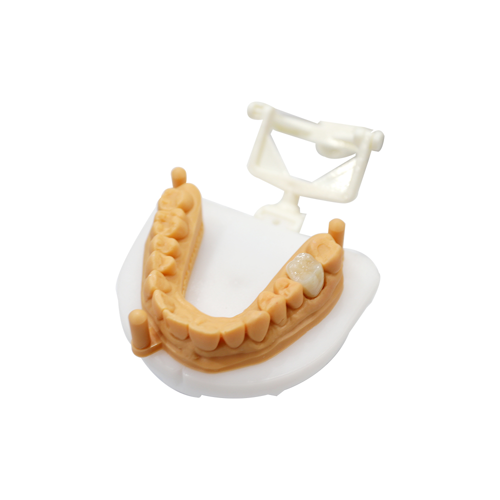


A 47-year-old male patient presented with a failing restoration on his lower right second molar. The tooth had undergone endodontic treatment two years prior, followed by a large composite buildup that fractured twice within eight months. His dentist referred the case to us with specific requirements for a durable, conservative restoration that could withstand high occlusal forces without requiring additional tooth reduction.
This case demonstrates our approach to posterior single-crown cases where clinical constraints—limited interocclusal space, high masticatory load, and compromised remaining tooth structure—demand careful material selection and design strategy.
Patient Information
Patient: 47-year-old male, non-smoker, no significant medical history. Bruxism suspected but not diagnosed.
Chief complaint: “The filling on my back tooth keeps breaking. I need something that will last.”
Clinical History
- Endodontic treatment completed 24 months prior (tooth #31 / lower right second molar)
- Initial composite buildup placed immediately post-endo; fractured at 4 months
- Second composite buildup (larger, reinforced with fiber post); fractured at 8 months
- Patient reports heavy chewing forces on the right side; opposing tooth is natural enamel
- No history of previous crown failures on other teeth
Clinical Examination (from scan and photos)
Remaining tooth structure:
- Buccal wall intact, approximately 3.5 mm height above CEJ
- Lingual wall partially intact, 2–3 mm height
- Mesial and distal walls compromised; interproximal contacts maintained by adjacent teeth
- No subgingival caries; margins cleanly prepared at or slightly above gingival level
Occlusal clearance: 1.3 mm at functional cusp; 1.5 mm at central fossa (measured from digital scan)
Opposing dentition: Natural mandibular second molar, moderate wear facets, no restorations
Interproximal contacts: Tight contact mesially with first molar; normal contact distally with third molar (present and functional)
Soft tissue: Healthy, no inflammation; probing depths within normal limits
Why This Case Was Challenging
- Minimal occlusal clearance (1.3 mm) ruled out layered zirconia or PFM; monolithic material required
- History of repeated fractures suggested either material inadequacy or occlusal overload—design had to address both
- Limited axial wall height (2–3 mm lingually) increased retention risk and stress concentration at margins
Why This Case Required Monolithic Zirconia
The patient’s history told us this wasn’t a simple crown case.
Two composite buildups had failed in twelve months. That’s not material failure—that’s a loading problem. Zirconia can be fabricated thin enough to hold up to maximum chewing forces up to 900 N, but only if the design accounts for where that load concentrates. This patient’s pattern (preferential right-side chewing, suspected bruxism) put him at the high end of ordinary loading.
Layered zirconia wasn’t an option. Veneer porcelain fractures under cyclic loading when occlusal clearance drops below 1.5 mm. Practitioner case reports consistently show veneer fractures at 18–36 months in similar cases, even with proper support geometry. The 1.3 mm clearance we had didn’t give us room for a competent veneer layer.
PFM would have required an additional 0.3–0.5 mm reduction to accommodate metal and porcelain thickness. The referring dentist explicitly requested no further tooth reduction—the axial walls were already at minimum retention height.
Monolithic zirconia was the only material that could meet three simultaneous constraints: minimal thickness (0.8–1.0 mm occlusal), adequate fracture resistance at reduced thickness, and no delamination risk (no layered interface).
What the Doctor Specified
The referring doctor sent a detailed case note with the digital scan:
| Requirement | Doctor’s Note | Clinical Rationale |
| No additional reduction | “Walls are at minimum height—cannot reduce further without exposing post” | Lingual wall 2–3 mm; further reduction would compromise retention and risk post exposure |
| Occlusal anatomy | “Functional cusp must clear by at least 0.2 mm in excursions; DO NOT over-contour” | Patient has tight lateral guidance; over-contouring would create premature contact and accelerate wear on opposing natural tooth |
| Margin design | “All margins supragingival or equigingival—patient has excellent hygiene, wants to maintain it” | Subgingival margins risk cement retention and inflammation; patient preference for cleanability |
| Delivery timeline | “Patient traveling for work in 14 days—needs crown seated before departure” | Standard 10-day turnaround covers this; no expedited handling needed |
Molar Zirconia Crown Design and Fabrication
Material Selection
3Y-TZP monolithic zirconia, full-contour design.
- Flexural strength: adequate for 0.8–1.0 mm occlusal thickness in posterior loading zones
- Translucency: mid-range (HT grade); aesthetic demand minimal for second molar
- Margin thickness: 0.6 mm feather-edge capable; no die spacer interference with supragingival finish line
We do not use 5Y-TZP for load-bearing single crowns. Its lower flexural strength creates unacceptable risk in thin sections under cyclic posterior load.
Design Strategy
Our CAD designer worked from the following parameters:
Occlusal thickness:
- Functional cusp: 1.0 mm (minimum for 3Y-TZP under high posterior load)
- Non-functional cusps: 0.8 mm
- Central fossa: 1.2 mm (maximum available clearance used for thickness, not anatomy height)
Cusp anatomy: Anatomically reduced—functional cusp morphology flattened to 20° incline (vs. 30–35° natural). This sacrifices some chewing efficiency but distributes load over a larger surface area and reduces shear stress at the occlusal-axial line angle.
Margin design: 0.6 mm feather-edge circumferentially. Supragingival on buccal, lingual, and distal; equigingival on mesial (tissue drape from scan indicated slight tissue overgrowth mesially).
Contact points:
- Mesial: 4.2 mm² contact area (slightly broader than natural to stabilize first molar)
- Distal: 3.1 mm² contact area (normal)
- Both contacts placed at occlusal third; no food trap risk identified
Internal fit: 80 µm cement space uniformly; no occlusal contact on die spacer zone (verified in virtual articulation).
Occlusal Adjustment Protocol
We provided written occlusal guidance with the crown delivery:
At Seating (Before Cementation):
- Verify full seating with no rocking; confirm marginal fit with explorer
- Check interproximal contacts with floss—should require moderate force, no tearing
- Mark MIP contacts with articulating film (8 µm); adjust to even contact distribution across occlusal table
- Do not adjust functional cusp tip—reduction there drops thickness below safe minimum
After Cementation: 5. Re-mark MIP; verify no high contact from cement film thickness 6. Check lateral excursions—crown should have light contact in working side, no contact in balancing 7. Check protrusive—light contact acceptable; heavy contact requires adjustment on non-functional slopes only
Key principle: Adjust non-functional anatomy first. Functional cusp and central fossa are minimum-thickness zones and should be preserved unless absolutely necessary.
Quality Control: What We Verified Before Shipping
Every case goes through a four-point check before it leaves our facility:
- Margin integrity: 10× loupe inspection under angled light; no chipping, no overcontour beyond 0.1 mm
- Occlusal thickness verification: Digital calipers at functional cusp and fossa; must meet or exceed design spec (1.0 mm / 1.2 mm)
- Contact point verification: Crown seated on master die; adjacent dies positioned; floss resistance checked
- Surface finish: No grinding marks on occlusal surface; all adjustment marks polished out with rubber points and re-polished to original surface roughness
We photograph and dimensionally verify every crown against the design parameters before it ships. If any check fails, the case goes back to design or manufacturing—not to you.
Try-In and Clinical Feedback
The crown was delivered within the 10-day window. Here is what happened at seating.
Patient Feedback
The patient’s first comment after trying the crown in — before cementation, while still checking the bite with articulating paper — was: “It doesn’t feel like a new tooth. It just feels like my tooth.”
That is the outcome we aim for with a posterior crown. A second molar is not a display tooth; the patient should stop noticing it.
What he reported during and after try-in:
- Comfort on closure: No sensation of the tooth being “too big” or “too tall.” He was checking whether it would catch on the opposing molar — it did not.
- Language awareness: Confirmed nothing was caught between his teeth when speaking. This is a common early-warning sign that contact points are too broad.
- Tongue comfort: The lingual contour against the adjacent molar felt natural — no bulk sensation.
- On chewing: He said the bite felt even and “solid,” with no preference shifting to the other side.
After seating and cementation (reported at the 2-week follow-up):
- No sensitivity to cold or hot. This is expected with a properly seated endodontically treated tooth, and it is one of the things we ask the doctor to confirm at follow-up.
- No pain on biting.
- No awareness of the crown during normal eating, including on the right side — which is where he previously concentrated his chewing after the buildups failed.
- His closing comment: “I’ve stopped thinking about that tooth. That’s how I know it’s working.”
Doctor’s Clinical Assessment
The referring dentist’s notes from seating and from the two-week review:
At seating (same day):
- Marginal fit: Fully seated with finger pressure, no rocking, no cement gap visible. “Seating was uneventful — this is the part where thin-section crowns often fight me.”
- Margins: Supragingival on buccal and lingual were fully visible and cleansable. The equigingival mesial margin was tight but accessible with a probe.
- Proximal contacts: Tight mesially, normal distally. Floss passed with the resistance expected on a properly contoured contact.
- Marginal integrity: No visible chipping at the feather-edge, including at the mesial equigingival margin — the location the doctor had flagged as the highest risk during planning.
- Occlusal adjustment required: Minimal. Two contacts were adjusted on non-functional slopes. The functional cusp tip was untouched, consistent with our design note.
- Excursions: Working-side contact confirmed; no balancing contacts; protrusive contact light and shared across adjacent teeth.
At the two-week review:
- Occlusal stability: No new wear facets. Contacts remained as delivered.
- Margins: No cement washout, no inflammation, probing depths unchanged.
- Composite restorations elsewhere in the patient: Intact.
- The doctor’s note: “Patient reports no awareness of the tooth. Given that this is his third restoration on this tooth in two years, I would normally counsel expectance. That advice felt unnecessary here.”
Why We Share This
We publish try-in feedback because it is the part of a case that is easy to claim and hard to fake. “It fits” is a measurement. “It feels like his own tooth” is a report from the person wearing it, and “I no longer counsel expectance on this tooth” is a clinical judgement made by a practitioner six weeks after placement.
We will still tell you when a case does not resolve this cleanly. What we will not do is present a case narrative where every step goes perfectly and the reader has no way to tell the difference between a real outcome and a marketing paragraph.
Zirconia Crown Thickness and Occlusal Clearance
We see constrained posterior clearance often enough that it is worth addressing directly, because it is where cases most often go wrong.
Below 1.5 mm functional cusp clearance: A layered zirconia or feldspathic/metal-ceramic restoration is a poor choice. The veneer layer needs bulk it will not have. Full-contour monolithic zirconia can be designed to 0.8–1.0 mm.
Below 0.8 mm functional cusp clearance: We flag this to the doctor before milling. It does not automatically mean the case cannot be done, but it changes the conversation. Options include adjusting the opposing tooth, changing the restoration design to a different material, or accepting a split (two-piece) restoration. We do not silently design a crown below the minimum thickness threshold to complete a case.
Clearance above 1.5 mm: A layered restoration becomes viable, and aesthetic demand plays a role. In most posterior single crowns we still recommend full-contour zirconia for posterior molars where aesthetics are not a driver — the mechanical profile is more predictable and there is no interface to fail.
Quality Standards We Work To
On material and process: ISO 13485 certified quality management system. FDA-listed custom medical device manufacturer (registration is not approval, and we do not describe it as approval). EU MDR compliant for custom devices.
On documentation: Material batch traceability, case number linked to every fabrication step, and a case sheet recording design parameters at each stage. If you need this for your records or for a patient conversation, it exists.
On communication: Design changes that affect the case — a modification to margins, a thickness that lands outside spec, a contact point we had to compromise — get told to you the day we find them, not at delivery.
Common Questions
Can I send a case with limited occlusal clearance?
Yes, and we will tell you the clearance situation before you commit to a design. Send the scan. We will measure it and come back with what is achievable in the space you have. If a layered restoration is not viable, you will know that in the first response rather than after the case is milled.
Do you use 3Y-TZP or 5Y-TZP for posterior crowns?
3Y-TZP for load-bearing posterior single crowns. 5Y-TZP trades flexural strength for translucency, and we do not want that trade in a molar. If a case is in an anterior position where translucency matters, the material choice changes.
Do you place abutment structures or come back to you for that?
Send what you have. If the case needs a core build-up, a fiber post, or anything else to be done intraorally before the final impression, we will tell you at the planning stage so you are not surprised at the delivery appointment.
What about endodontically treated teeth — do you need anything special?
Flag the endodontic status and the date of treatment on the case sheet. For a molar crown the relevant factors are ferrule and residual wall height, both of which affect retention. We design the margin and taper to your recorded preparation, but the final retention strategy is your clinical call.
How long does a case take?
Standard turnaround is 10 working days from receipt of a complete digital case. If your patient has a travel date or an event, tell us on the case sheet and we will confirm what is realistic against that date rather than you finding out late.
What if the crown does not seat?
Send us a photo and the seating response. The usual causes are debris in the preparation, a mis-recorded margin, or a scanner artefact on the preparation. We will tell you which one it is. If it is ours, we re-mill and re-send; we do not send you a crown that fits its own die and fails in the mouth.
Do you work with labs as well as clinics?
Yes. Send cases the same way — the communication channel and the case sheet are identical. If you are working an intraoral scan or a model, tell us which and we will confirm the workflow.
Order a Molar Zirconia Crown from ASD Dental Lab
Send the digital case and we will come back with clearance measurements, a design direction, and a realistic delivery date against your patient’s timeline.
What to include:
- Intraoral scan or model, with the prepared tooth and adjacent teeth captured
- Case number, tooth number, and shade
- Material preference if you have one
- Endodontic status and date, if applicable
- Margin type and whether you are bonding or cementing
- Patient’s delivery deadline, if there is one
If you would rather talk it through before sending, tell us what the case looks like and we will tell you whether it is one we would take.