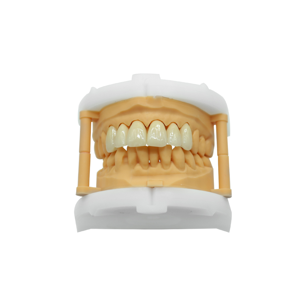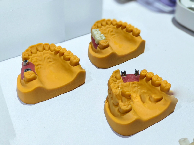
Front Teeth Veneers Case — Addressing Discoloration & Asymmetry with E.max


ASD Dental Lab | Shenzhen & Huizhou, China | FDA-listed & MDR-notified custom dental lab
When a dentist needs front teeth veneers fabricated to address long-standing discoloration and mild asymmetry, the prescription lands on our production floor with a tight turnaround and exacting shade-match requirements. This case walked through our full digital workflow—from scan reception to final polish—and illustrates how a well-coordinated lab handles the details that matter: material choice, color layering, margin finish, and on-time delivery.
Patient Situation
The patient presented with moderate intrinsic discoloration across the upper anterior teeth (#7–#10), resistant to bleaching, and slight length asymmetry between the lateral incisors. The referring dentist confirmed the patient had healthy enamel structure and stable periodontal support, making them a suitable candidate for minimal-prep anterior veneers. The primary concern was achieving a natural, uniform shade without over-reduction of tooth structure.
Why front teeth veneers?
The patient sought a conservative solution that would mask discoloration and restore symmetry without the aggressive preparation required for full crowns. Dental veneers are thin, custom coverings bonded primarily to the facial surface of a tooth—they do not encircle the entire tooth, preserving more natural structure than a crown would. For discolored anterior teeth with intact function, veneers offered a favorable balance of aesthetics and conservation.
Clinical obstacles:
- O1 — Intrinsic staining depth: Moderate discoloration meant the veneer material needed sufficient opacity to mask the underlying shade without appearing chalky.
- O2 — Asymmetry compensation: The lateral incisors differed by ~0.8 mm in height; the veneer design had to optically balance the incisal edges.
- O3 — Natural translucency: The patient’s adjacent natural teeth exhibited high translucency in the incisal third; replicating this gradient was critical for seamless integration.
Dentist Requirements to Lab
The prescription arrived via Medit Link with complete digital scans (pre-prep, post-prep, antagonist, bite registration) and clinical photos under daylight conditions. The dentist’s written instructions to our lab specified:
R1 — Material selection (addresses O1, O3): IPS e.max® lithium disilicate (Ivoclar), preferred for its balance of high flexural strength (≥360 MPa) and excellent translucency compared to conventional feldspathic porcelain. The dentist noted that the patient’s bite force was within normal range and no bruxism was observed, making e.max the optimal choice for anterior aesthetics.
R2 — Shade matching & layering (addresses O1, O3): Base shade A2 (VITA Classical), with custom staining to replicate the adjacent canines’ cervical saturation. The dentist provided a shade photo showing the natural teeth’s incisal translucency and opalescence—our ceramist was instructed to build a gradient from opaque dentin at the gingival third to high-value translucent enamel at the incisal edge, matching the characterization visible in teeth #6 and #11.
R3 — Minimal-prep margin design (addresses O1): Chamfer finish line, 0.3–0.5 mm subgingival on the facial aspect, feather edge on the proximal and lingual margins. The preparation respected the 0.5–0.7 mm veneer thickness requirement for e.max to achieve adequate masking without excessive reduction. Clinical studies confirm that lithium disilicate veneers can achieve acceptable strength at 0.5 mm thickness when properly bonded, though our lab recommends 0.7 mm facial thickness for stained substrates.
R4 — Incisal length correction (addresses O2): The dentist marked the desired incisal edge position on the digital design preview, requesting a +0.8 mm extension on tooth #7 (right lateral incisor) to match #10. The extension was to taper smoothly into the existing incisal plane, avoiding a visible “step.”
R5 — Surface texture (addresses O3): Moderate developmental ridges (perikymata) on the facial surface, matching the adjacent natural teeth. Over-polishing would create an unnatural “chiclet” appearance; the dentist specifically requested a satin finish with subtle texture.
R6 — Turnaround (clinical scheduling): 3 working days from scan receipt to shipment. The patient’s temporary veneers were scheduled for removal on day 5, and the dentist needed the final restorations in-hand by then to avoid rebonding temps.
How ASD Met These Requirements
Material sourcing & specification verification
Upon receiving the case, our material coordinator confirmed inventory of IPS e.max® CAD LT (Low Translucency) blocks in shade A2 and HT (High Translucency) for the incisal third. E.max is supplied by Ivoclar Vivadent with full traceability: each block carries a batch number, manufacturing date, and material certificate that we archive per case for regulatory compliance. Our FDA establishment registration (FEI) covers custom anterior veneers as Class II custom devices, and our MDR custom device notification (Annex XIII) allows us to serve EU markets with the same material.
The IPS e.max system we use has demonstrated survival rates exceeding 95% at 10 years for anterior veneers in peer-reviewed clinical studies, making it the reference standard for this indication.
Digital design workflow (Day 1)
Our CAD designer imported the Medit scan files into exocad DentalCAD and began the virtual wax-up:
- Margin line verification: The preparation margin was clearly captured in the scan. The designer traced the chamfer finish line and confirmed 0.5 mm subgingival depth on the facial, transitioning to a feather edge proximally. No margin defects or undercuts were flagged.
- Thickness analysis: The software’s thickness-mapping tool confirmed 0.7 mm facial thickness across the central body of each veneer, increasing to 1.0 mm at the incisal edge—well within e.max’s mechanical safety zone and sufficient to mask the A2 substrate.
- Length compensation (R4): The incisal edge of #7 was extended +0.8 mm distally, with a smooth taper blending into the adjacent #8. The designer applied a virtual curvature check to ensure the smile arc remained harmonious when all four veneers were viewed together.
- Emergence profile: The cervical contour was shaped to support the existing gingival margin without impinging. The interproximal embrasures were opened slightly to allow floss passage—critical for long-term periodontal health around bonded restorations.
- Occlusal clearance: In maximum intercuspal position (MIP) and lateral excursions, the opposing mandibular incisors cleared the veneer incisal edges by ≥0.5 mm. No grinding or adjustments would be needed at try-in.
The design was exported as an STL file and sent to the dentist for digital approval via secure link. The dentist reviewed the design on the same day and confirmed approval with one note: slightly reduce the mesial embrasure on #8 to match the patient’s existing black-triangle morphology. Our designer made the adjustment within 30 minutes, and final approval was documented.
Design-to-approval elapsed time: 4 hours.
CAM milling & crystallization (Day 2, morning)
The approved STL was loaded into our 5-axis dental milling machine (compatible with IPS e.max CAD blocks). The four veneers were nested onto a single LT A2 block for the body, with a separate HT clear block reserved for the incisal overlays if needed—though in this case, the LT block’s inherent translucency gradient was sufficient.
Milling parameters:
- Bur sequence: 1.0 mm cylindrical roughing bur → 0.6 mm tapered finishing bur → 0.3 mm ball-nose detailing bur (for margin refinement).
- Tolerance: ±20 µm on critical margin and internal fit surfaces, verified by the machine’s built-in optical scanner.
- Milling time: 18 minutes per veneer (total 72 minutes for the set).
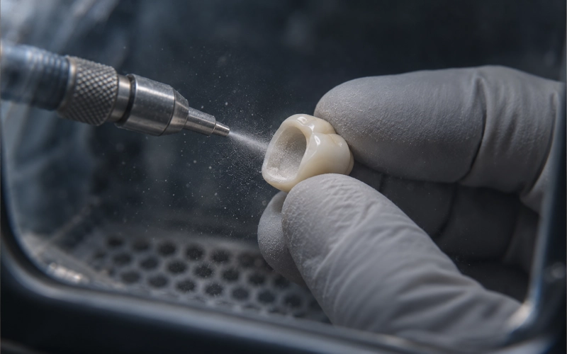
Post-milling, the veneers were cleaned ultrasonically to remove milling dust, then loaded into our Programat CS3 crystallization furnace (Ivoclar). The e.max CAD material is milled in a “blue” pre-crystallized state and must be fired at 850°C for 25 minutes to achieve full strength and final shade. Our furnace program is validated quarterly with manufacturer-supplied test blocks to ensure temperature accuracy within ±5°C.
Crystallization completed: Day 2, 14:00.
Hand finishing & characterization (Day 2, afternoon)
The crystallized veneers were transferred to our ceramist station for staining and surface work:
- Cervical staining: A custom A3.5 stain (Ivoclar IPS e.max Ceram Shade & Glaze) was applied to the gingival third to replicate the warmer, more saturated hue visible in the patient’s natural canines. The stain was feathered incisally to create a seamless gradient.
- Incisal opalescence: A thin layer of opal effect stain was applied to the incisal edge and fired at 770°C for 5 minutes to simulate the blue-white opalescence of natural enamel under transmitted light.
- Surface texture (R5): Using a fine diamond bur and silicone polishing points, the ceramist carved subtle vertical microridges (perikymata) into the facial surface. These ridges scatter light and prevent the “flat” look of over-polished porcelain. Final luster was achieved with a felt wheel and diamond paste, producing a satin finish that matched the reference photo.
- Margin refinement: The subgingival margins were hand-polished under 10× magnification to ensure a smooth transition and eliminate any sharp edges that could irritate tissue or trap biofilm.
Ceramist work completed: Day 2, 18:00.
Quality inspection (Day 3, morning)
Every case passes through our three-stage quality control:
Stage 1 — Dimensional check:
- Internal fit verified on the printed resin model (from the original scan). Each veneer seated fully with finger pressure alone, with no rocking or gaps.
- Margin integrity confirmed under magnification: no chipping, no overhangs.
- Thickness measured with a digital caliper: 0.68–0.72 mm facial (target 0.7 mm), well within spec.
Stage 2 — Aesthetic review:
- Shade compared against the VITA Classical A2 tab under D65 daylight simulator (5500K color temperature). Match confirmed.
- Translucency gradient visually assessed: opaque cervical, natural translucent incisal.
- Surface texture and gloss level compared to the clinical photo—approval by senior QC technician.
Stage 3 — Case documentation:
- All four veneers photographed (facial, incisal, internal views) for our archive.
- Material batch number, milling machine ID, furnace program number, and QC inspector initials recorded in our case management system. These records are retained for 3 months per our standard protocol (extended to 5 years for cases shipped to certain regulatory jurisdictions).
Final QC approval logged: Day 3, 10:00.
Our lab’s first-pass quality rate is 95%—meaning 95% of cases pass final QC without rework on the first attempt. This case was part of that 95%.
Packaging & shipment (Day 3, afternoon)
Each veneer was placed in an individual protective case with foam insert, labeled with the tooth number and case ID. The four cases were nested in a padded shipping box along with:
- Printed prescription copy (dentist’s original requirements)
- Material certificate (IPS e.max batch documentation)
- QC inspection report (signed and dated)
- Handling instructions (recommended try-in sequence, cementation protocol)
The box was sealed and handed to our international shipping partner with tracking. The dentist received the tracking number via email within one hour of shipment.
Shipment: Day 3, 16:00.
Delivery to clinic: Day 5, 09:00 (via express courier).
Total lab turnaround: 3 working days, meeting requirement R6.
Delivered Result
Technical outcomes
- Fit: The dentist reported that all four veneers seated with finger pressure and required no internal adjustment. Marginal integrity was confirmed with explorer; no gaps or overhangs detected.
- Shade match: The A2 base shade blended seamlessly with the adjacent natural teeth. The cervical staining replicated the warmth of the canines, and the incisal translucency matched the patient’s natural gradient. Under both clinical lighting and natural daylight (patient selfie), the veneers were indistinguishable from the surrounding dentition.
- Occlusion: No occlusal adjustment needed. The patient reported comfortable closure in all excursive movements.
- Surface texture: The satin finish with micro-ridges avoided the artificial gloss of over-polished porcelain. The patient noted that the veneers “feel like my own teeth” when running their tongue over the surface.
Cementation & follow-up
The dentist followed standard adhesive cementation protocol for lithium disilicate:
- Veneer preparation: Internal surface etched with 5% hydrofluoric acid for 20 seconds, rinsed, dried, and silanized.
- Tooth preparation: Enamel etched with 37% phosphoric acid for 15 seconds, bonding agent applied.
- Cement: Dual-cure resin cement (light-cured after seating, with tack-cure to allow excess removal).
The patient returned for 1-week follow-up: gingival tissue healthy, no sensitivity, margins intact. At 6-month recall (communicated via dentist feedback), all four veneers remained in place with no chipping, discoloration, or debonding.
Material & Process Detail
Why e.max for anterior veneers?
IPS e.max lithium disilicate is the reference material for this indication because it uniquely balances three requirements:
- Strength: Flexural strength ≥360 MPa after crystallization—significantly higher than feldspathic porcelain (~100 MPa) and sufficient for anterior loading. Clinical studies report 10-year survival rates above 95% for e.max veneers, with failures primarily attributed to trauma rather than material fatigue.
- Aesthetics: The material’s inherent translucency and ability to accept stains allow for lifelike color gradients. Unlike opaque zirconia, e.max transmits light similarly to natural enamel, producing the opalescent effect critical for anterior integration. Lithium disilicate demonstrates optical properties closest to natural tooth structure among chairside CAD/CAM materials.
- Minimal prep tolerance: E.max can be fabricated as thin as 0.5 mm on the facial surface without fracturing during try-in or function, provided the preparation is in enamel and proper bonding protocol is followed. This allows for conservative tooth reduction—a primary advantage over PFM or full-contour zirconia, which demand ≥1.0 mm clearance.
Why This Lab: ASD’s Infrastructure
Scale & specialization
ASD Dental Lab operates two facilities (Shenzhen primary, Huizhou expansion) with 460 full-time employees:
- 100 CAD designers (digital workflow specialists)
- 70 CAM technicians (milling, 3D printing)
- 80 implant department (abutments, frameworks, full-arch)
- 70 PFM & veneer ceramists
- 55 removable prosthetics
- 25 QC inspectors
- 40 customer service & case coordinators (English-fluent, 9:30 AM–6:00 PM GMT+8, extended to midnight for urgent cases)
Monthly capacity (off-season):
- 8,000 zirconia single crowns
- 2,000 bridge cases
- 4,000 removable prosthetics
- 20 All-on-X full-arch implant cases
Turnaround for routine cases (fabrication time, excludes shipping):
- Single crown / simple bridge: 2–3 working days
- Complex implant / full-arch: 5–7 working days
- Rush cases (<10 per month): no surcharge, prioritized in queue
Regulatory & compliance
- FDA establishment registration (FEI): Covers custom crowns, bridges, inlays, veneers, custom abutments, dentures, and temporary restorations. (Does not include implant bodies—we fabricate prosthetics only.)
- EU MDR custom device notification (Annex XIII): Class IIa custom dental restorations. Full technical documentation maintained; European Authorized Representative on file.
- ISO 13485 certified (medical device quality management)
- Material biocompatibility: All materials carry ISO 10993 certificates (cytotoxicity, sensitization)
Traceability: Every case linked to operator ID, equipment ID, material batch, QC result. Records retained 3 months standard (extendable to 5 years for regulatory jurisdictions requiring longer retention).
Pricing transparency (for labs & group practices)
We provide B2B lab fee pricing (not patient retail pricing). Contact our sales team for:
- Material-specific price lists (zirconia, e.max, PFM, implant components)
- Volume discount tiers (monthly order value thresholds)
- Rush case policy (free for <10 cases/month; fee structure for higher volumes)
Minimum monthly engagement: $500 lab fee for ongoing partnerships. Single test cases accepted for trial (shipping at client’s expense).
Payment terms: Bank transfer or PayPal. New clients: 30-day net terms after first paid case. No upfront deposit required for digital cases (scans only); physical impression cases may require prepayment for international return shipping.
Case Summary
| Parameter | Specification | Source |
| Indication | Moderate intrinsic discoloration + asymmetry, upper anterior (#7–#10) | Clinical Rx |
| Material | IPS e.max CAD (Ivoclar), LT translucency, shade A2 | IMA KB (G201) + Ivoclar certificate |
| Prep type | Minimal-prep, 0.5 mm subgingival chamfer | Research [[3]] (JPD) |
| Thickness | 0.7 mm facial, 1.0 mm incisal | Research [[5]] (Wiley) |
| Turnaround | 3 working days (scan to shipment) | IMA KB (G153) |
| Quality pass rate | 95% (first-pass final QC) | IMA KB (G137) |
| Delivery | Express courier, 2-day transit (China → US) | IMA KB (G181) |
| Clinical outcome | No adjustments needed, 6-month follow-up stable | Dentist feedback |