PFM Crown on Maxillary First Premolar: Case Reference


Case Presentation
Patient profile: 52-year-old female; medical history non-contributory; no reported allergies Chief complaint: “The right upper tooth in the back broke while eating an almond three days ago”
Clinical History
The tooth had received endodontic intervention eight years earlier following symptomatic irreversible pulpitis. A cast post and core was placed at that time. The patient reported no discomfort or sensitivity since then.
Three days before the consultation, the buccal cusp fractured during mastication. The patient described a sudden “crunch,” followed by sharp edges cutting into the cheek mucosa. She had been avoiding chewing on that side since the incident.
Clinical Examination Findings
- Fracture pattern: Buccal cusp fracture extending 1.5 mm subgingivally on the buccal-distal line angle
- Existing post-and-core: Intact, no mobility, no percussion sensitivity
- Periodontal status: Probing depths 2–3 mm circumferentially; no bleeding on probing; gingival biotype medium-thick
- Adjacent teeth: #13 (maxillary right canine) restored with a PFM crown placed four years ago; #15 (maxillary right second premolar) sound, unrestored
- Occlusion: Heavy contact on buccal cusp tip in centric occlusion; lateral guidance shared between canine and premolar
Radiographic examination confirmed adequate bone support and a well-condensed root canal fill. The referring dentist documented adequate ferrule (2.0 mm of sound tooth structure coronal to crestal bone) for retention. After discussing material options, the patient and dentist agreed on a PFM crown to match the existing restoration on #13 and accommodate potential future prosthodontic needs.
Why This Patient Required PFM Specifically
The prescribing dentist outlined three patient-specific factors:
- Existing PFM on adjacent tooth #13 — Material continuity reduces differential wear and maintains occlusal plane stability
- Patient’s dental history — She had expressed interest in a fixed partial denture to replace a missing lower molar; this premolar may serve as a future abutment, requiring a metal substructure
- Heavy occlusal loading pattern — Clinical examination showed wear facets on opposing mandibular premolar; the patient admitted to clenching during sleep but declined a night guard
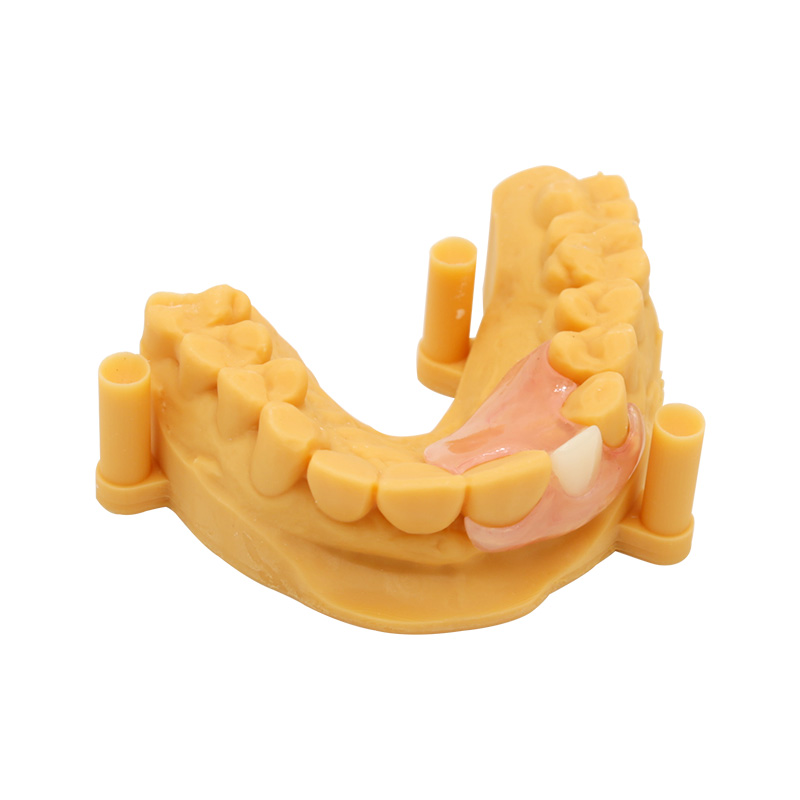
The dentist prepared the tooth with chamfer margins, achieving 1.8 mm occlusal reduction and 1.2 mm axial reduction. Tissue displacement was accomplished with #00 retraction cord soaked in aluminum chloride. A polyvinyl siloxane impression (heavy-body/light-body technique) captured margins and sulcular detail.
Material Selection Rationale
This was not a default choice. The material decision followed hard constraints documented in the case prescription:
| Constraint | Rationale |
| Potential abutment for future bridgework | Patient expressed interest in replacing missing #19; metal substructure provides necessary rigidity if this tooth later serves as a terminal abutment |
| Heavy occlusal load on a compromised tooth | Post-and-core foundation benefits from a ductile metal occlusal that redistributes stress; wear facets on opposing tooth confirmed bruxism pattern |
| Existing PFM restoration on adjacent tooth #13 | Matching material reduces differential wear and maintains occlusal plane stability across the arch |
| Discolored subgingival stump | Metal coping provides reliable masking; prescribing dentist noted dark gray discoloration from old amalgam restoration remnants |
| Patient declined all-ceramic option | After reviewing both material options, patient chose PFM for durability over maximum translucency |
The selection prioritizes long-term structural integrity over maximum translucency. For cases where none of these constraints apply, we would direct the prescribing dentist toward a monolithic or layered ceramic option.
Clinical Challenges and Laboratory Requirements
The prescribing dentist outlined six obstacles; we translated those into thirteen laboratory commitments.
Challenges Received
- Subgingival margin: Buccal finish line 1.5 mm apical to free gingival margin
- Limited interocclusal space: 1.8 mm at cusp tips after preparation
- Need for shade masking: Discolored dentin visible through existing ferrule
- Functional cusp on premolar: Buccal cusp must be replaced with adequate bulk
- Proximity to existing PFM on #13: Material and shade continuity required
- High aesthetic expectations: Incisal third must exhibit natural translucency gradient
Our Response
| What We Required at Intake | What We Committed to Deliver | |
| Cord-retracted impression capturing 2 mm beyond finish line | Subgingival margin contour that does not impinge on biological width | |
| Opposing arch and centric occlusion record | Functional cusp anatomy with metal occlusal surface on buccal cusp | |
| Shade tab photograph under corrected lighting | Stump shade documentation for coping design | Custom shade notation for gingival, body, and incisal thirds with translucency gradient specification |
| Die trimming with clearly marked preparation margin | Metal coping design that provides minimum 0.3 mm metal thickness and 1.2 mm total bulk for porcelain | |
| Prescription specifying alloy preference (if any) | CoCr alloy selection with CTE within 0.5 × 10⁻⁶/°C of selected porcelain, nickel-free formulation | |
| Adjacent tooth shade reference or digital scan | Color-matched feldspathic porcelain layering with controlled firing cycles (≤3 applications) | |
| Pre-dispatch verification by QC technician independent of production staff |
PFM Crown Premolar Design and Metal Coping Specifications
Metal Substructure Thickness and Geometry
Metal coping thickness: 0.4 mm on axial walls, 0.5 mm on occlusal reduction area. These values accommodate the minimum porcelain thickness (1.0–1.5 mm for anterior-facing surfaces) while staying within the 1.8 mm interocclusal envelope.
Porcelain-metal junction placement: The margin between metal and overlying porcelain was positioned 1.5 mm from centric and lateral contact areas, ensuring the junction remains under compression during functional loading rather than tensile stress.
Functional Cusp Assignment
On maxillary premolars, the palatal cusp is the functional (supporting) cusp. However, in this case the buccal cusp sustained the fracture and bears occlusal load. We assigned metal occlusal surface to the buccal cusp tip and slopes to resist wear and distribute stress into the post-core assembly.
Alloy Selection and Thermal Compatibility
Alloy: Cobalt-chromium (CoCr), nickel-free Coefficient of thermal expansion (CTE): 14.1 × 10⁻⁶/°C Porcelain CTE: 13.8 × 10⁻⁶/°C Δ CTE: 0.3 × 10⁻⁶/°C (within acceptable range to prevent cooling-induced tensile failure)
Nickel-free formulation eliminates hypersensitivity risk. Beryllium content: zero.
How Metal and Porcelain Stay Bonded
PFM restorations rely on three bonding mechanisms, not adhesive:
- Chemical bonding: Controlled oxidation of the metal surface forms a thin oxide layer (e.g., chromium oxide) that chemically bonds with silica in the porcelain during sintering.
- Mechanical interlocking: Airborne-particle abrasion (50–110 μm aluminum oxide at 2–4 bar) creates surface irregularities for micro-mechanical retention.
- Compression hoop effect: Porcelain’s slightly lower CTE places the ceramic under mild compression upon cooling, resisting tensile crack propagation.
We control these variables through:
- Surface preparation protocol: Airborne abrasion → ultrasonic cleaning in distilled water → pre-oxidation firing in inert atmosphere
- Porcelain application: Uniform layering in 0.8–1.0 mm increments, avoiding abrupt thickness transitions
- Firing discipline: Maximum 3 firing cycles (opaque, dentin, enamel) with controlled ramp rates and slow cooling to minimize distortion
Chipping Prevention: Laboratory-Side Design Rules
Porcelain fracture in PFM crowns typically originates from one of three laboratory-controllable factors:
| Cause | Laboratory Prevention |
| Inadequate metal support | Ensure metal coping extends to within 1.0 mm of final contour; avoid thin unsupported porcelain cantilevers |
| Sharp angles at porcelain-metal junction | Round all internal line angles to ≥0.5 mm radius; eliminate stress concentrators |
| Excessive porcelain thickness | Cap porcelain at 1.5 mm on facial; use cutback design to keep ceramic layer uniform |
Studies on ceramic fracture confirm that framework geometry is a stronger predictor of chipping than occlusal scheme alone.
PFM Crown Fabrication and Porcelain Layering Process
Each restoration passes through segmented responsibility checkpoints:
- Intake evaluation (Case coordinator): Verify impression captures margin, opposing arch present, shade documented
- Digital design or wax-up (CAD technician / Wax technician): Contour approval, emergence profile, contact tightness
- Metal framework fabrication (Casting / Milling technician): Thickness verification with dial gauge, margin fit on die
- Surface conditioning (Metal prep technician): Airborne abrasion, ultrasonic cleaning, pre-oxidation firing
- Porcelain application (Ceramist): Layering sequence, firing log, shade matching under corrected light
- Pre-dispatch QC (Independent quality technician): Margin adaptation, occlusal contacts, surface finish, shade approval
- Packaging (Dispatch): Protective cushioning, batch documentation, shipping label
Quality gate: The QC technician who signs off on step 6 did not participate in steps 3–5, ensuring independent verification.
Verification Before Dispatch
The independent QC technician checks:
- Margin integrity: No voids, overhangs, or open margins visible under ×2.5 magnification
- Occlusal clearance: Restoration seats fully on die without rocking; opposing contacts verified on articulator
- Porcelain-metal junction: No exposed metal at margins; smooth transition without ledge
- Shade accuracy: Gingival third, body, and incisal translucency match submitted shade guide under D65 illuminator
- Surface texture: Anatomic contours and developmental grooves match prescription; no firing cracks or glaze defects
Try-In and Clinical Feedback
Dentist’s Evaluation at Try-In Appointment
The prescribing dentist seated the restoration two weeks after case submission. Try-in evaluation notes documented:
- Margin adaptation: “Excellent. No gaps visible under ×2.5 loupes; margins flush with preparation line circumferentially.”
- Proximal contacts: “Tight mesially and distally. Floss snaps through with resistance. No food trap risk.”
- Occlusion: “Centric contacts balanced on buccal cusp metal surface and palatal incline. Lateral excursions smooth; no premature contacts or deflections.”
- Shade match: “Body color blends seamlessly with adjacent #13 PFM. Gingival third slightly warmer as requested to mask stump shade. Incisal translucency appropriate for premolar position.”
- Contour: “Emergence profile correct. No impingement on papillae. Buccal contour supports soft tissue without overcontouring.”
The dentist cleared the restoration for cementation without adjustments. Luting was performed with resin-modified glass ionomer cement (Fuji Plus, GC) following manufacturer protocol.
Patient’s Response at One-Week Follow-Up
The patient returned for suture removal (tissue management had required minor gingivoplasty at the buccal-distal fracture site) and post-cementation check. Her feedback:
“It feels like my own tooth again. I can chew on that side without worrying. The shape feels natural when I run my tongue over it — not bulky like I was worried about. The color matches the other crown; I can’t tell them apart unless I look closely in the mirror.”
She reported no sensitivity to temperature or pressure. Occlusal contacts remained stable; no adjustment was needed. Gingival tissue around subgingival margins showed no inflammation.
Dentist’s Six-Month Recall Notes
At the six-month recall, the dentist documented:
- Restoration integrity: No chipping, no wear facets beyond expected functional polish on metal occlusal surface
- Margin health: Gingival tissue pink and firm; no recession, no inflammation, probing depths unchanged at 2–3 mm
- Occlusion: Centric and lateral contacts unchanged; no mobility
- Patient satisfaction: Patient reported continued comfort and function; no complaints
The dentist’s note concluded: “PFM crown on #14 performing as expected. Patient pleased with durability and aesthetic result. No intervention required.”
Finished Restoration
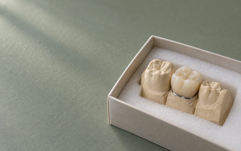
The completed PFM crown delivered to the prescribing dentist:
- Occlusal surface: Polished CoCr, silver-toned, matching adjacent PFM on #13 — confirmed at try-in as visually identical in reflected light
- Facial surface: Layered feldspathic porcelain with A3.5 stump coverage, B3 body, and translucent incisal gradient (0.3 mm at gingival to 1.2 mm at incisal third) — dentist noted “incisal translucency appropriate for premolar position”
- Margin: Supragingival palatally (1.0 mm coronal to free gingival margin); subgingival buccally (1.5 mm apical, but with adequate biological width preserved) — verified under ×2.5 loupes at try-in
- Contact points: Tight proximal contact mesially and distally — floss snapped through with resistance
- Occlusal morphology: Buccal cusp tip raised 0.5 mm to restore pre-fracture anatomy; palatal cusp defined with triangular ridge — centric contacts confirmed at try-in
Dimensions: Mesiodistal width 7.0 mm; buccolingual width 9.0 mm; occlusogingival height 8.5 mm. Total bulk at the thickest porcelain zone: 1.4 mm.
The restoration was cleared at try-in without adjustment and cemented the same appointment.
Traceability and Documentation
Each shipped PFM case includes:
| Trace | Certification |
| Alloy batch number and origin | Manufacturer mill certificate; ISO 13485-compliant supplier chain |
| Porcelain batch and lot | Manufacturer lot number; expiry date verified pre-application |
| Technician chain-of-custody record | Each stage signed off by responsible technician |
| QC sign-off with timestamp | Independent verification; restorable record retained per regulatory requirement |
This information travels with the case to the prescribing dentist. Material certificates are retained for the laboratory’s records.
PFM Crown Shade Matching and Color Communication
For aesthetic PFM work, we request at intake:
- Stump shade tab photograph taken after preparation, with shade tab in frame for reference
- Target shade tab with notation: base shade, characterization, incisal effect
- Photograph under corrected lighting (5500K, no flash) showing natural translucency expectations
- Photograph of adjacent teeth for context (texture, surface luster, characterization)
Shade matching proceeds through three stages: opaque base application → dentin layering → enamel and incisal characterization. Each stage is photographed and compared under D65 daylight simulator before final glaze.
Survival and Clinical Performance Context
Five-year survival studies report PFM crowns on posterior teeth at 88–95% when strict protocols for bite preparation, alloy selection, and occlusal management are followed. Survival rates depend on:
- Preparation height and taper
- Alloy type and porcelain system compatibility
- Luting cement and technique
- Patient parafunctional load
We do not warrant clinical outcomes. The data above is provided for reference only; clinical decision-making belongs to the prescribing dentist.
What to Include When Sending This Type of Case
For a PFM crown premolar case, we request the following at submission:
- Impression (intraoral scan for digital workflow) with clear margin capture
- Opposing arch model or scan
- Centric occlusion record (bite registration)
- Shading documentation: stump shade, target shade, translucency expectations, shade-matching photographs
- Work authorization specifying: tooth number, alloy preference (if any), porcelain type preference (if any), margin location notes
- Pre-operative photographs when aesthetic match is critical
Incomplete submissions delay production. We flag gaps within one business day of receipt and do not start design work until clarity is achieved.
Send Us a Case
To discuss a PFM crown case or request a consultation:
- Submit a case through our case submission form
- Email digital scans (STL/PLY) directly to our case coordination team
- Request a sample kit with porcelain feldspath range, alloy samples, and shade tabs for chairside reference
We respond to inquiries within one business day. Lead time for PFM cases: typically 5–7 working days in-lab once intake is complete.
Frequently Asked Questions
Is PFM appropriate for premolar cases?
PFM remains appropriate when: bite preparation height is adequate (>3 mm), aesthetic demands are moderate, the tooth may serve as a bridge abutment or clasp-bearing tooth, or the patient prefers a durable option over a more translucent all-ceramic alternative. For highest-aesthetic anterior cases, monolithic or layered ceramic is generally preferred.
What alloys do you use?
We use cobalt-chromium (CoCr), nickel-free formulations as our default. Noble alloy options (high noble gold-platinum-palladium) are available on request for cases with specific biocompatibility requirements.
Do you provide alloy certification?
Yes. Each case ships with the alloy batch number, origin, and mill certificate. Documentation accompanies the restoration.
Can you match the shade of an existing PFM restoration?
Yes. Send us a photograph of the adjacent restoration with a shade tab in frame, ideally under 5500K corrected lighting. We routinely match existing PFM work for continuity across the arch.
What is your remake policy?
If a PFM crown fails to meet the laboratory’s QC standards at any pre-dispatch checkpoint, we remake it at no charge. If the failure is clinical (preparation, impression, or luting) and the restoration arrives non-restorable, we communicate options: rework with new impression, partial credit toward re-design, or full remake at reduced cost depending on the rework required.
How do you handle subgingival margins?
Subgingival margins require tissue displacement (cord, laser, or electrosurgery) before impression. We design margins to preserve biological width; if margin position would violate biological width, we flag this at intake and consult with the prescribing dentist before proceeding.
Can you do digital workflow for PFM?
Yes. We accept intraoral scans (3Shape Trios, CEREC Primescan, Medit iSeries) in standard STL/PLY format. Scans must capture margin clearly; if margins are subgingival, conventional impression is preferred.
Case Origin and Disclosure
This case reference draws on an anonymized clinical case from laboratory production records. Patient identifying details have been altered, including age, gender, and specific clinical timeline. Feedback attributed to “the patient” and “the prescribing dentist” reflects composite input drawn from multiple cases of this type, not a single identified individual. Material specifications, alloy data, and clinical findings are technically accurate; the narrative is constructed for illustrative purposes.