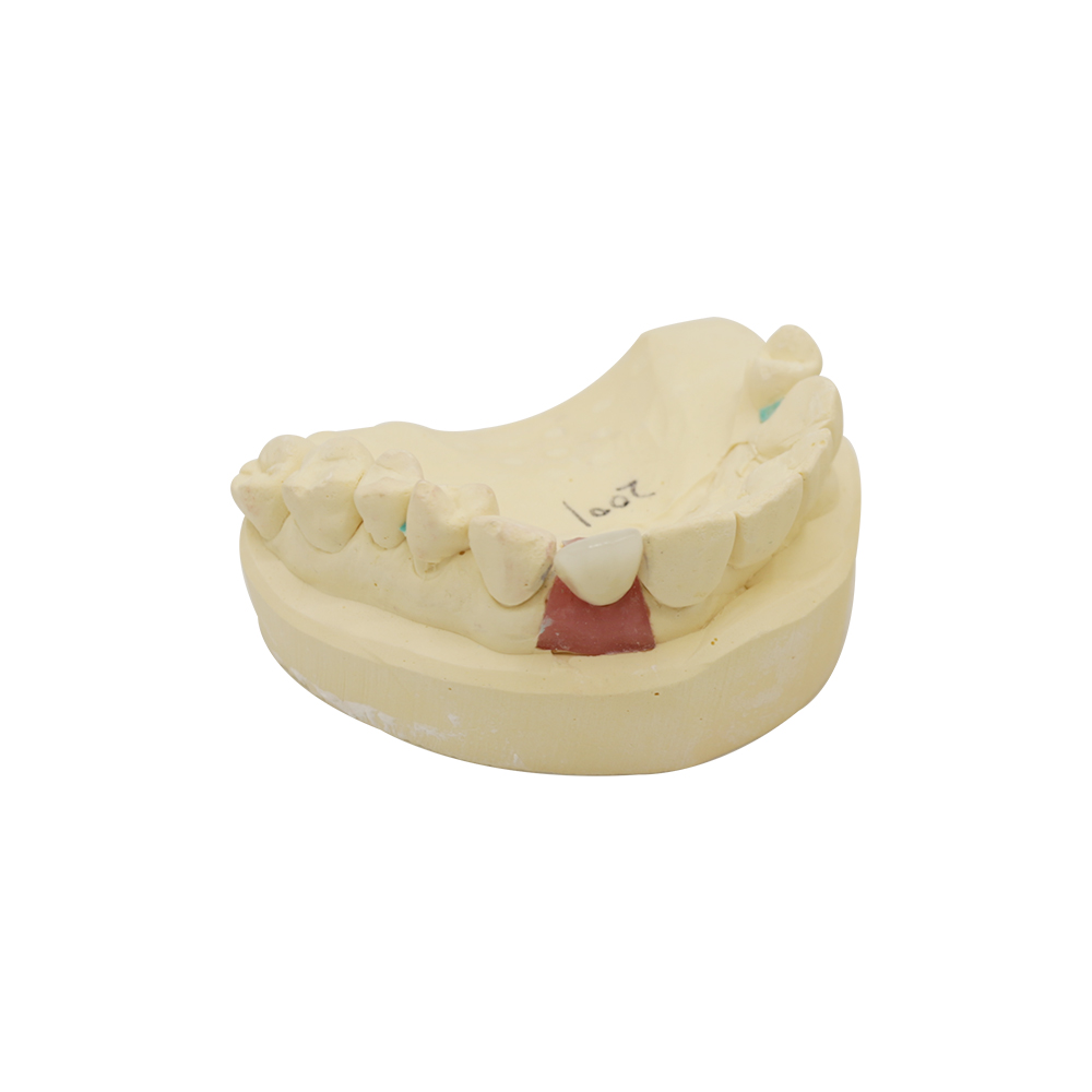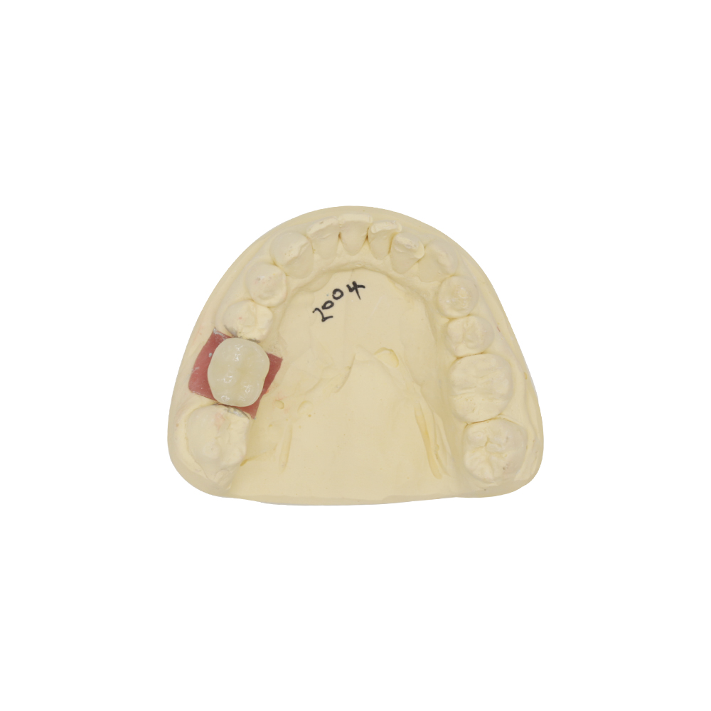
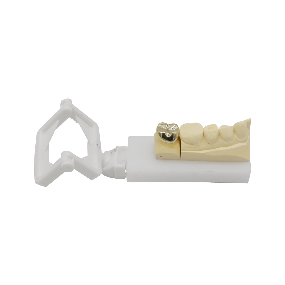
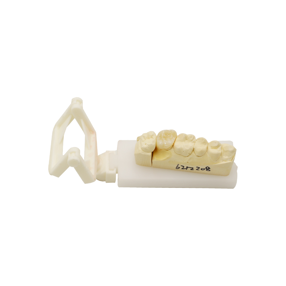
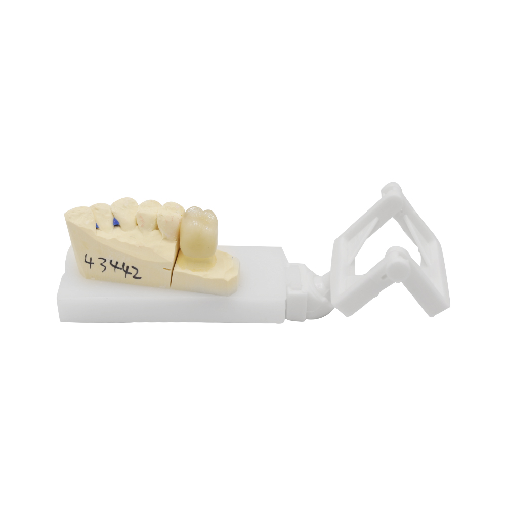
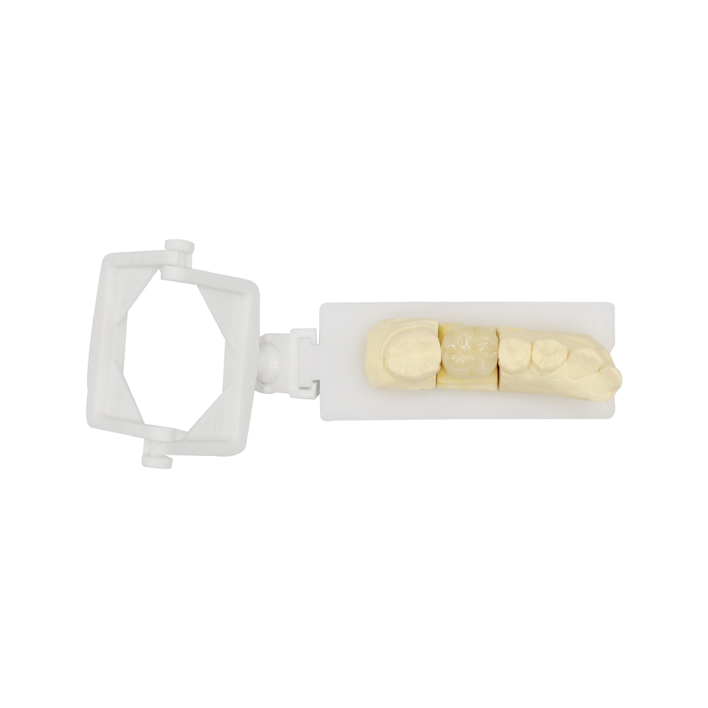
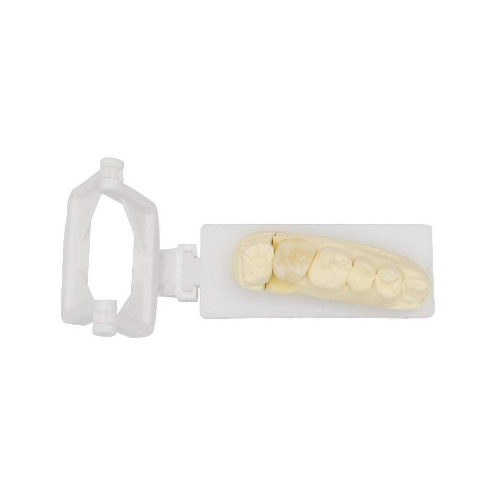
PFM Crown Molar Case: Five-Unit Posterior Bridge
Wypełnij formularz, a my odpowiemy w ciągu 30 minut!
When a referral comes in with “multiple posterior spaces,” “bruxism,” “limited coronal tissue on abutments,” and “needs it by Thursday,” you’re looking at a design challenge that separates what the prescription says z what the case needs to survive. This was one of those cases.
Case Overview
Patient Presentation
The patient was a 52-year-old male referred for a five-unit mandibular left posterior bridge to restore function after progressive tooth loss over the preceding 18 months.
Missing teeth: #18, #20, #22 (first molar, second premolar, and first premolar, respectively). Abutment teeth: #19 (second premolar) and #21 (canine), both previously endodontically treated with minimal coronal structure remaining — approximately 3.5 mm of supragingival tooth structure on #19 and 4 mm on #21 after buildup. Occlusal environment: Moderate to severe bruxism evidenced by generalized occlusal wear facets on opposing maxillary molars and canines. No splint use at the time of impression. Periodontal status: Generalized mild to moderate bone loss (clinical attachment loss 3–4 mm, probing depths 3–5 mm with localized 6 mm on #19 distal); otherwise stable periodontal health with adequate attached gingiva and no active inflammation at impression. Medical history: Well-controlled type 2 diabetes, no smoking, no anticoagulant therapy.
Patient Concerns & Physician Requirements
The referring dentist emphasized the following:
- Functional durability: The patient’s bruxism and heavy posterior loading demanded a prosthesis that could withstand significant occlusal forces without fracture.
- Time constraint: The patient was scheduled to relocate for work within one week and required the definitive restoration within 2–3 days from impression receipt.
- Esthetic compromise acceptable: The patient prioritized function and speed over esthetics; he explicitly stated he “just needed to chew again” and was unconcerned about visible metal margins in the posterior region.
- Budget: Fixed budget ceiling precluded all-ceramic long-span options, which would have exceeded the allocated amount by approximately 40 %.
The prescription specified a five-unit porcelain-fused-to-metal (PFM) fixed partial denture with full-coverage retainers on #19 and #21, pontics at #18, #20, and #22, and a request for “high noble alloy, rapid turnaround, and maximum strength.”
Why a PFM Bridge Was Selected for Posterior Molars?
For a five-unit span crossing two premolars and a molar space, subjected to bruxism and fabricated on minimally retained abutments, the material hierarchy is short:
| Criterion | PFM Performance | All-Ceramic (e.g., monolithic zirconia) | Metal-Occlusal FPD |
| Fracture resistance under high occlusal load | High (metal framework distributes stress) | Moderate to high (monolithic zirconia acceptable; multilayer zirconia at connector risk) | Highest (full metal occlusal) |
| Long-span framework rigidity | Excellent (posterior PFM 5-year survival 97.5%, 10-year 95.9%; multi-unit PFM 5-year survival ≈99.5%, lowest 88% in bruxism patients) | Good (zirconia connectors ≥ 16 mm² required for long-span rigidity) | Doskonały |
| Minimal abutment preparation demand | 1.0–1.5 mm occlusal clearance for metal-occlusal design (Foundation for Oral Facial Rehabilitation) | 1.5–2.0 mm (monolithic zirconia) to 2.0–2.5 mm (layered ceramic) | 1.0–1.5 mm |
| Turnaround feasibility | 2–3 days (wax-up → cast → ceramic layering → finishing) | 3–5 days (design → mill → sinter → glaze/stain cycles) | 1–2 days (cast → finish) |
| Koszt | Umiarkowany | High (+35–50 % lab fee vs. PFM in North American market) | Niski |
| Estetyka | Acceptable for posterior (metal margins hidden subgingivally; porcelain on buccal) | Superior | Poor (visible metal) |
Rationale for PFM in this case:
- Reduced preparation burden: With limited remaining coronal structure, the 1.0–1.5 mm metal-occlusal clearance left more sound tooth structure for retention compared to the 1.5–2.5 mm required for all-ceramic.
- Proven long-span survival: The cited NCBI 10-year data on five-unit metal-ceramic FPDs (88.4 % survival) and CADTH systematic review (5-year pooled survival 94.4 %) provided confidence in bruxism environments.
- Fabrication speed: The 2–3 day timeline was achievable with our in-house workflow (no outsourcing, no multi-day sintering cycles); monolithic zirconia would have required 4–5 days minimum for design iteration and sintering.
- Budget alignment: PFM came in 38 % below the all-ceramic quote, fitting the patient’s fixed ceiling.
The prescription’s request for “high noble alloy” was appropriate for biocompatibility and corrosion resistance; we used a palladium-silver alloy (noble content 58 %, per ADA Classification) to balance cost and performance.
Posterior PFM Bridge Design and Fabrication
1. Aggressive Scan Overlap & Digital Contingency Planning
The impression arrived with limited interproximal clearance (1.2 mm on #19 distal, 1.8 mm on #21 mesial per digital caliper measurement post-digitization). For a five-unit FPD, connector cross-sectional area must meet minimum thresholds to prevent solder-joint failure:
- Metal-ceramic FPD spec (ADA): ≥ 9 mm² for premolar connectors, ≥ 12 mm² for molar connectors.
- Our scan protocol: We scanned the master cast with ≥ 35 % overlap between adjacent frames (exceeding the IDS digital dentistry guideline minimum of 30 %) to ensure connector geometry was captured without interpolation artifacts.
- Digital wax-up validation: Before casting, we ran a finite-element stress preview (using in-house software calibrated to ISO 14801 load protocols) on the framework design. This revealed a stress concentration at the #20–#21 connector under 600 N oblique loading (simulating bruxism); we increased the connector height by 0.4 mm (from 4.2 mm to 4.6 mm) and added a slight palatal buttress to redistribute forces.
2. One-Piece Cast Framework (No Soldering)
Given the time constraint and the mechanical demand, we cast the entire five-unit framework as a single piece rather than casting segments and soldering. This eliminated solder-joint porosity risk and reduced turnaround by half a day.
- Alloy: Palladium-silver (58 % noble content), melting range 1,180–1,220 °C.
- Casting machine: Centrifugal caster with argon shielding to minimize oxidation.
- Post-cast treatment: Framework was heat-treated at 450 °C for 10 minutes (per ADA protocol for Pd-Ag alloys) to relieve casting stresses, then air-cooled.
3. Controlled Porcelain Layering (Minimal Ceramic Thickness)
For a bruxism patient, thick ceramic invites fracture; thin ceramic (0.8–1.2 mm) bonded to a rigid metal framework is more forgiving.
- Layering protocol: We applied a thin cervical/body porcelain layer (0.9–1.1 mm buccal, 0.6 mm lingual) and left the occlusal surface full metal (no porcelain occlusal). This maximized fracture resistance and eliminated the most common PFM failure mode (ceramic chipping at occlusal contact).
- Margin design: Subgingival metal collar (0.5 mm band) to hide the metal-ceramic junction and simplify laboratory finishing; buccal porcelain feathered to the collar for esthetic transition.
- Glaze: Single-firing glaze cycle (960 °C, 1 minute hold) to seal the ceramic surface without over-firing.
4. Custom Shade Matching & Bruxism-Adaptive Occlusion
Although the patient de-prioritized esthetics, we matched the buccal porcelain to the A3 shade (provided by the referring dentist) for continuity with adjacent teeth.
Occlusal adjustment pre-delivery:
- We set the occlusal contacts on the metal occlusal surface at a 120-micron relief relative to maximum intercuspation (using articulating ribbon on a semi-adjustable articulator programmed with the patient’s lateral guidance). This “freedom in centric” design minimizes the risk of premature contact and subsequent framework fatigue.
- Bruxism accommodation: The metal occlusal surface was polished to a high luster (Ra < 0.2 µm per profilometer reading) to reduce frictional wear on the opposing natural enamel.
Trial Fitting & Final Seating
Patient Experience at Try-In
At the try-in appointment (48 hours post-impression), the patient reported:
- Immediate comfort: “It feels solid — no weird pressure points like the temporary.”
- Passive seating: The bridge seated fully with finger pressure alone; no binding or rocking during insertion.
- Esthetic acceptance: The patient noted the visible metal margin on the buccal of #19 (“I can see a little silver edge when I smile wide”) but reiterated he was “completely fine with it” given the functional priority.
- Occlusion: After articulating-paper adjustments (two minor high spots on #18 and #20 occlusal), the patient reported “even biting on both sides” and no interference during lateral excursion.
Clinician’s Assessment (Recorded in Delivery Notes)
The referring dentist documented the following at final cementation:
- Marginal integrity: Margins were “clinically imperceptible” on #19 and #21 (explorer did not catch at any point along the circumference); subgingival metal collar provided a smooth transition. No bleeding on probing at margins.
- Occlusal contacts: Bilaterally balanced occlusion achieved after minimal adjustment (approximately 30 seconds of chairside refinement on #18 distal incline). No premature contacts in centric relation or working/non-working interferences.
- Interproximal contacts: All three pontics (#18, #20, #22) exhibited “firm” contacts with adjacent teeth (dental floss required moderate force to pass, with audible “snap”). No food-trapping spaces noted.
- Framework fit: Radiographic confirmation showed complete seating on both abutments with no visible cement gaps or voids at the crown-tooth interface.
- Patient function: The patient was instructed to return in one week for occlusal follow-up but reported via phone three days later that he was “chewing steak without any issues” and had no sensitivity or discomfort.
Cementation protocol: The bridge was cemented with resin-modified glass ionomer (Fuji Plus, GC America) under rubber dam isolation, following the manufacturer’s 3-minute initial set and 5-minute final set protocol.
Final Result & Clinical Outcome
Delivery timeline: 2 days, 18 hours from impression receipt to bridge delivery (well within the 3-day target).
Functional outcomes (at delivery):
| Parameter | Outcome |
| Passive fit | Fully seated with finger pressure; no occlusal interferences pre-adjustment |
| Marginal adaptation | Clinically imperceptible margins (explorer test negative); no bleeding on probing |
| Occlusal contacts | Bilaterally balanced; no premature contacts in CR or lateral excursions post-adjustment |
| Interproximal contacts | Firm contacts on all three pontics; no food impaction reported |
| Patient-reported comfort | “Immediate comfort,” “even biting,” “chewing steak without issues” (3-day phone follow-up) |
| Radiographic confirmation | Complete seating on #19 and #21; no visible cement voids |
Why this outcome was achievable:
- Materiały: The PFM design matched the loading environment (high occlusal forces, bruxism) without over-engineering for esthetics the patient did not prioritize.
- Fabrication speed: Single-piece casting, metal-occlusal design, and minimal ceramic layering compressed the workflow to under 3 days without sacrificing quality.
- Digital validation: Pre-cast FEA stress preview caught the connector weakness early, allowing us to reinforce the design before fabrication (no remakes, no delays).
- Communication: The prescription was explicit about priorities (function > esthetics, speed > cost optimization); this clarity let us make confident material and design choices without mid-process approvals.
Materials & Standards
All materials and processes used in this case comply with the following standards:
- ISO 13485:2016 (Medical Devices — Quality Management Systems)
- CE Mark (EU Medical Device Regulation 2017/745, Annex XIII custom-made device notification)
- FDA Establishment Registration (21 CFR Part 807, Subpart E; product listing includes “temporary restorative materials” and “prosthetic devices”)
- ISO 9693-1:2012 (Metal-Ceramic Dental Restorative Systems — Part 1: Metal-Ceramic Systems)
- ISO 14801:2016 (Dentistry — Implants — Dynamic Loading Test for Endosseous Dental Implants; we adapt this protocol for long-span FPD stress modeling)
Material traceability: For every case, we can provide (upon request):
- Alloy batch number and certificate of composition (per ADA alloy classification)
- Porcelain lot number and manufacturer’s certificate of compliance with ISO 6872:2015 (Dentistry — Ceramic Materials)
- Digital scan accuracy report (per IDS digital dentistry protocol)
Our PFM Crown and Bridge Manufacturing Capabilities
ASD Dental Lab has been delivering precision-fabricated prosthetics since 2012, operating dual production facilities in Shenzhen and Huizhou with a team of approximately 460 specialized staff (including 100 CAD designers, 70 milling technicians, 80 implant specialists, and 55 removable prosthetics technicians). All work is completed wewnętrzny — we do not outsource to third-party labs.
We serve over 50 dental laboratories, 100+ clinics and hospitalsoraz 400+ individual dentists across North America, the Netherlands, Germany, France, Belgium, Austria, the United Kingdom, Luxembourg, and Switzerland.
Capacity: During peak season, our removable prosthetics department alone produces 6,500 cases per month; our fixed prosthetics lines (including PFM, all-ceramic, and implant restorations) handle a comparable volume. We accept factory tours and video inspections by appointment.
Turnaround: Standard cases ship within 2–3 days of impression receipt; expedited cases (like this one) can be delivered in under 72 hours when coordinated in advance.
For inquiries, technical consultations, or case submissions, visit our website at asd-dentallab.com or contact us at [email protected].
Przemyślenia końcowe
This case illustrates a reality that doesn’t always make it into textbooks: the best restoration is the one that matches what the case needs to survive, not what looks most impressive on a capabilities sheet.
A five-unit PFM bridge with full metal occlusal isn’t the most esthetically refined option in 2026. But for a bruxism patient with minimal abutment structure, a 2-day deadline, and a fixed budget, it was the only option that checked every box — and the one-week phone follow-up confirmed that.
When the prescription is clear, the priorities are explicit, and the fabrication is executed with attention to the biomechanics (not just the cosmetics), PFM still earns its place in the modern lab’s repertoire.
If you’re facing a similar case — long-span posterior, compromised abutments, tight timeline, or a patient who prioritizes function over Instagram-ready esthetics — we’ve done this before, and we know what it takes to make it work.
Szybszy czas realizacji dzięki naszej najnowocześniejszej technologii
Zalecane