PFM Bridge Anterior — Case Reference


🕢 [ ¡Rellene el formulario y le responderemos en 30 minutos! ]
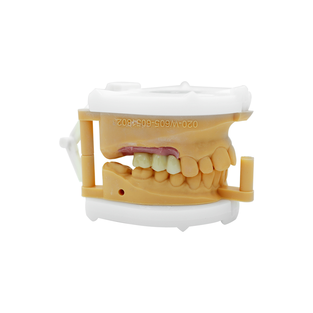
Long-span anterior PFM bridge over a resorbed ridge, in a patient for whom implants were ruled out
Case at a Glance
| Patient | Adult, mid-30s |
| Chief zone | Mandibular anterior (FDI 31–36 region) |
| Missing units | Multiple lower anteriors, replaced for years by an all-acrylic removable partial denture |
| Ridge condition | Narrow, reduced vertical height, following a failed ridge augmentation |
| Occlusal risk | Untreated nocturnal bruxism; night guard declined |
| Restoration | Tooth-supported porcelain-fused-to-metal bridge |
| Abutments | Both canines (sound, minimal existing buccal restorations) plus one premolar (existing resin crown) |
| Core difficulty | Long span, aesthetic zone, limited clearance and high load in one case |
Patient Presentation
The presenting complaint. He was tired of taking his denture out to eat. The lower anterior segment had been missing for roughly a decade and had been replaced with an all-acrylic removable partial denture: a plate he removed to eat, that visibly moved, and that he was conscious of every time he smiled. He wanted something fixed. He also wanted it to look like his own teeth rather than like a prosthesis. Those were his two requirements and he stated them plainly at the first appointment.
History. Some years earlier, a ridge augmentation had been carried out in the lower anterior region with the intention of placing implants. The graft did not hold. It resorbed, leaving a ridge narrow buccolingually and short in height. A second grafting procedure was discussed and the patient declined it.
That single decision shaped the entire case. Bone volume was insufficient and further grafting was off the table, so implants were not available. What remained was a tooth-supported fixed bridge, and every design question after that followed from it.
Findings.
- Both canines were sound. The left carried minimal existing buccal restorations, essentially intact coronal structure with no large failing restoration driving the decision.
- The right premolar adjacent to the span already carried a resin crown. It was serviceable, and the clinician was keen to avoid re-preparing it further.
- The edentulous ridge was narrow with reduced vertical height, and the soft tissue over it was thin. This ruled out anything needing a labial flange or a bulky pontic form.
- Anterior clearance was limited. The opposing dentition had intruded into the functional space, so the vertical room for a metal core plus porcelain was already tight, and in places very tight.
- No radiographic or clinical evidence of active periapical or periodontal pathology on any proposed abutment.
The habit nobody had addressed. He ground his teeth at night. It was in the history and visible in the wear patterns. A night guard was recommended and declined. That absence removed the protective cushion that would otherwise have absorbed a good share of the cyclic load the bridge was about to take, which made it a design input rather than a footnote.
His goal in his own terms. A fixed restoration, natural-looking in the front, that he could eat with and then forget about. Nothing more was asked of him than that.
Why an Anterior PFM Bridge Was Selected
With implants excluded and the patient unwilling to continue with a removable appliance, a tooth-supported fixed bridge was the only fixed option. That made the framework the governing consideration, because in a long span the substructure rather than the veneering material decides whether the restoration survives. A metal framework carries the load and allows a small, controlled amount of flex across the span, with porcelain layered on for appearance.
The published multi-unit data supports that reasoning. A systematic review of tooth-supported fixed prostheses reported five-year survival of approximately 99.5% for porcelain-fused-to-metal against approximately 92% for all-ceramic systems, and a large single-crown cohort reported five-year anterior survival above 96% with veneer chipping under 2%. Sources are listed at the foot of this page. Material selection remains the treating clinician’s decision in every case.
The second reason was repairability. A metal-ceramic restoration that chips can usually be repaired at the surface. An all-ceramic alternative that fails at a connector generally does not come back.
What the Clinician Asked the Lab to Do
Every instruction traced back to one of the constraints above. This was the brief as received:
| Constraint identified | Instruction to the laboratory |
| Long span over a resorbed ridge | PFM framework in a high-strength alloy; connectors sized and reinforced for the span; load shared evenly across the abutments; connector dimensions recorded and documented. |
| Untreated bruxism | Minimise exposed porcelain in function, using metal occlusal and lingual surfaces wherever clearance allows; uniform, controlled porcelain thickness; verify metal substructure thickness antes de layering begins. |
| Limited anterior clearance | Keep total occlusal thickness within the available space; no over-contoured occlusal surfaces; confirm seating and clearance on the die before proceeding. |
| Narrow ridge, thin soft tissue | Pontic design with adequate ridge clearance and hygiene access; no tissue-trapping contacts; keep pontics clear of excursive contact. |
| Aesthetic zone | Porcelain-shoulder margin positioned to minimise visible metal at the gingival line; shade matched to the supplied photographs with characterisation across the anterior span; framework try-in required before final porcelain firing. |
| Conservative abutment intent | Restrict the abutment scheme to the agreed teeth; do not enlarge the preparation beyond the prepared records supplied. |
| Records and verification | IOS/STL (or PVS) upper and lower, opposing arch, bite registration and shade photographs; a verification jig for the bridge; a dedicated framework try-in stage in the workflow. |
Anterior PFM Bridge Design and Fabrication
Framework design. The bridge was CAD-designed as a single metal substructure in an alloy selected for flexural strength and metal-ceramic bonding performance. Given the span and the lost vertical ridge height, connector geometry was calculated rather than drawn by rule of thumb, and the dimensions were recorded against the case file so they could be checked at try-in rather than judged by eye.
Substructure verified before porcelain. The metal was measured after casting and before any veneering porcelain went on. Where clearance was restricted, this is the step that decides whether the porcelain has room to sit at a workable thickness. Too little metal and the veneer ends up thin and vulnerable. Too much metal and the occlusal surface has to be over-reduced to compensate. Both failures show up here, cheaply, rather than after layering.
Margin design. A porcelain shoulder was placed on the labial surfaces across the aesthetic span so that no metal line would show at the gingival margin. This was the most consequential aesthetic decision in the case. His complaint had been about how the old restoration looked, and a visible grey margin would have reproduced exactly the problem he was trying to escape.
Occlusal metal where clearance ran short. Rather than thinning porcelain into a space that could not support it, metal was carried through on the occlusal and lingual surfaces at the points where vertical room ran out, and the pontics were kept clear of excursive contact.
Framework try-in before firing. The framework went back for trial seating before the final porcelain and glaze. Fit, seating and occlusion were confirmed at that stage with the clinician present, so any correction could be made to metal, which is the point at which correction is still straightforward.
Records retained. Alloy composition, batch number, processing parameters and the verification measurements were logged against the case. Our facility runs an ISO 13485-based quality management system and holds CE marking, and documentation is retained so that any individual case can be reconstructed later.
PFM Bridge Framework Try-In and Feedbac
As this is a representative Case Reference, what follows reflects the typical feedback pattern for this configuration rather than a verbatim patient record.
At the framework try-in. With the metal seated, the clinician checked abutment fit and the relationship of the pontics to the ridge before anything was layered. The specific concern at this stage was hygiene access under the pontics. Against a thin, resorbed ridge, a pontic that contacts the tissue too broadly is hard for the patient to clean and tends to become sore. Adjustments were made at metal stage, where that is straightforward, rather than after glazing.
After seating. Once the bridge was placed and the occlusion settled, two things were checked that often go unrecorded and matter more than appearance:
- Whether the ridge stayed comfortable under the pontics over extended wear. He was asked about this specifically, before being asked about appearance.
- Whether the anterior guidance felt natural, or whether the new bridge had altered his bite.
What the patient reported. His first comment was that he no longer had to remove anything to eat, which after a decade of a removable plate was the actual point of the case. He said the front teeth felt like his own. He noticed the difference for the first few days and then stopped noticing it, which was the outcome he had been aiming at. He remained aware of the grinding habit, having been told plainly that it was a factor in how the bridge was loaded and that it was still unaddressed.
What the clinician reported. Fit and marginal adaptation were recorded as satisfactory at seating, with no pressure areas on the ridge and no occlusal interference in either excursion. Marginal and contact relationships were within acceptable limits on check, and the shade was accepted against the supplied photographs. The closing note recorded two advisories rather than reassurances: that the patient still needed counselling on the grinding habit and that a night guard should be revisited, and that the ridge tissues under the pontics should be reviewed at routine recalls so that early pressure or inflammation did not develop unnoticed.
We do not make medical claims. Material selection, restoration design, case suitability and every clinical decision remain with the treating clinician.
About the Lab
Established in 2012, we are a full-service dental laboratory manufacturing in Shenzhen and Huizhou, China. We are a direct production facility, not a trading intermediary. Roughly 460 people work a nine-stage in-house process from case intake and CAD design through CAM milling, porcelain layering, finishing, inspection and packing, with nothing sent out to third-party labs. We currently supply more than 50 laboratories, over 100 clinics and more than 400 dentists across the United States and Europe. Factory tours and audits are welcome at both sites.
Fuentes
- Sghaireen MG, et al. (2022). Evaluation and Comparison of Five-Year Survival of Tooth-Supported Porcelain Fused to Metal and All-Ceramic Multiple Unit Fixed Prostheses: A Systematic Review. Cureus, 14(10):e30338.
- Behr M, Zeman F, Baitinger T, Galler J, Koller M, Handel G, Rosentritt M (2014). The clinical performance of porcelain-fused-to-metal precious alloy single crowns: chipping, recurrent caries, periodontitis, and loss of retention. Revista Internacional de Prostodoncia, 27(2):153-160.
- ISO 13485:2016. Medical devices — Quality management systems — Requirements for regulatory purposes. International Organization for Standardization.
This Case Reference is provided for professional reference. Individual outcomes depend on patient factors, clinical technique, material selection and post-placement care. Clinicians remain responsible for diagnosis, treatment planning, material selection and informed consent.