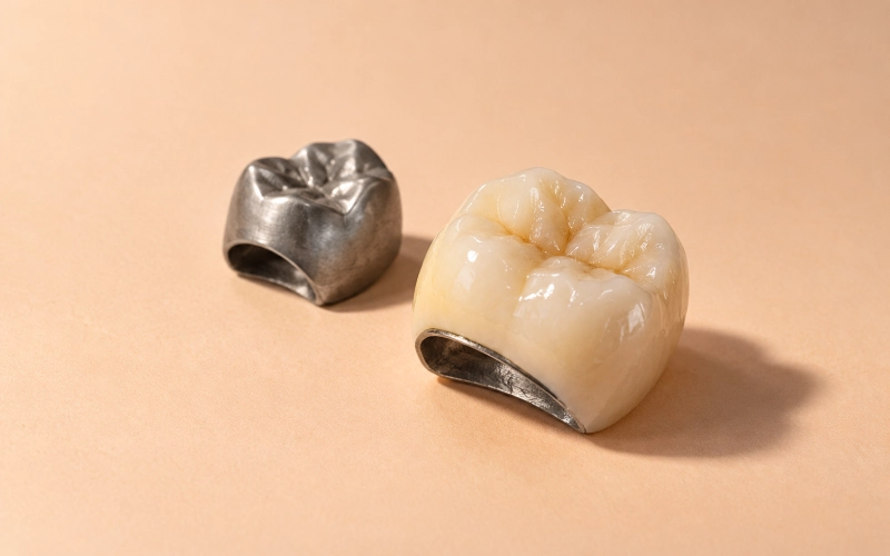
Temporary Molar Tooth Crown Case Study: Repeated Crown Fractures


Wypełnij formularz, a my odpowiemy w ciągu 30 minut!
The prescription slip from the referring practice read: “Fabricate temporary crown, tooth #31 (lower right second molar). Patient has fractured three previous crowns on this tooth. Deliberately position restoration out of occlusion to minimize further damage to remaining tooth structure. Patient is undecided between fourth permanent crown vs. extraction.”
This was not a routine interim restoration request. It was a diagnostic holding pattern disguised as a temporary crown—and the clinical question embedded in it was whether the tooth could be kept viable long enough for the patient to make an informed choice without destroying what was left of it in the process.
The Tooth’s History
Lower right second molar, tooth #31. The patient had root canal work performed on this tooth approximately nine years earlier. Since then:
- First permanent crown (PFM): fractured at the margin after 14 months
- Second permanent crown (full-contour zirconia): fractured mesial cusp after 22 months
- Third permanent crown (layered zirconia): fractured distal cusp and part of the occlusal table after 18 months
The patient reported a history of nocturnal bruxism and had been prescribed a night guard years ago, though compliance was inconsistent. At the time the case came to our lab, she told the dentist she was “afraid to chew on that side” and asked whether extraction might be the safer option. The referring dentist wanted to give her time to decide—but only if a temporary restoration could survive long enough to hold space and prevent drift without becoming the fourth failure.
Why the Case Came to Our Lab
The referring practice had sent the case to another lab first. That lab returned a standard bis-acryl temporary crown designed for normal occlusal contact. It fractured during try-in.
The practice then contacted us with a revised specification: “We need a temporary crown that will not break, even if that means it cannot function as a normal tooth. The patient needs time, not another failure.”
The key phrase in that request was “even if that means it cannot function.” The dentist was explicitly asking for a deliberate compromise: a restoration that would hold space and maintain soft-tissue contour, but intentionally kept out of occlusion to eliminate fracture risk. That is not a standard interim design—and it required us to document every decision so the patient and dentist could later evaluate whether the tooth’s structural integrity had been preserved or further compromised.
The Clinical Obstacles That Shaped Our Approach
Four factors made this case high-risk:
- Post-endodontic brittleness with insufficient ferrule. The referring dentist noted in the prescription that the remaining tooth structure above the gingival margin measured approximately 1.5–2 mm buccally and lingually, with near-zero ferrule on the mesial wall. The biomechanical literature consistently identifies insufficient ferrule as a primary risk factor for crown fracture in endodontically compromised molars.
- Documented high occlusal load history. Three permanent crowns fractured in sequence, all within 22 months or less. Even if the patient’s night-guard compliance improved, the cumulative load history suggested that any restoration in functional occlusion would carry unacceptable fracture risk during the interim period.
- Unclear structural prognosis. The patient was undecided between investing in a fourth permanent crown or accepting extraction. That meant the temporary restoration needed to function as a diagnostic tool: if the remaining tooth structure degraded further during the interim period (e.g., subgingival fracture, recurrent decay, apical pathology), that information would inform the patient’s final decision. We could not design a temporary crown that might mask or accelerate that degradation.
- Patient psychology and trust. The patient had already experienced three fractures. Another fracture—even of a temporary restoration—would likely push her toward extraction regardless of the tooth’s actual prognosis. The temporary crown had to survive not just biomechanically, but psychologically: it had to be the first restoration on that tooth that did not fail.
What the Dentist Specified (Seven Requirements)
The prescription included seven explicit requirements, which we interpreted and documented as follows:
R1. Deliberately position the occlusal surface 0.3–0.5 mm out of occlusion in maximum intercuspation (MIP).
↳ Verified via opposing-arch articulation and digital caliper measurement at try-in.
R2. Maintain proper mesial and distal contact with adjacent teeth (#30, #32) to prevent drift.
↳ Contact points documented at 0.05 mm mesial (tooth #30), 0.04 mm distal (tooth #32), measured post-mill with feeler gauge.
R3. Restore buccal and lingual contour to support soft tissue and facilitate hygiene access.
↳ Contour designed to match pre-fracture emergence profile, verified against pre-operative photos provided by the referring practice.
R4. Use a material that can survive months-long function in a high-bruxism environment—without occlusal contact.
↳ We selected milled PMMA (polymethyl methacrylate) rather than bis-acryl composite. PMMA has higher flexural strength (≥60 MPa per ISO 20795-1:2013 Type 1) and superior resistance to water sorption and discoloration over extended service periods.
R5. Design the restoration so it can be removed and replaced without further damaging the tooth structure.
↳ We used temporary cementation protocol documentation: the restoration was designed for cementation with non-eugenol temporary cement (e.g., Temp-Bond NE), and we provided the referring dentist with written guidance on controlled removal using a crown remover or ultrasonic scaler to minimize torsional stress on the remaining tooth structure.
R6. Provide contact and occlusal clearance documentation that the dentist can reference at follow-up appointments.
↳ We included a printed specification sheet listing mesial/distal contact measurements, occlusal clearance at six points (mesiobuccal, distobuccal, mesiolingual, distolingual, central fossa, distal marginal ridge), and photographic documentation of the pre-cementation margin fit.
R7. If the temporary crown survives the interim period without fracture or debonding, that survival data will help the patient decide whether to proceed with a fourth permanent restoration.
↳ This was the referring dentist’s exact wording. It made clear that the temporary crown was not just a placeholder—it was part of the diagnostic workup.
Temporary Molar Crown Design and PMMA Fabrication
This case required three fabrication steps that went beyond our standard interim-crown workflow:
Step 1: Opposing-arch data acquisition and occlusal clearance mapping.
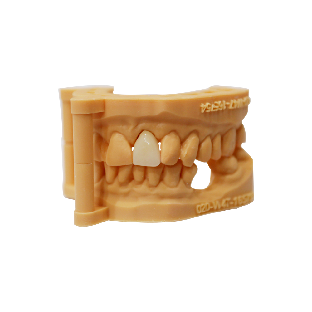
We requested and received a stone model of the opposing arch (maxillary right quadrant) and a bite registration in MIP. We mounted both models on a semi-adjustable articulator and mapped the occlusal clearance at six points on the #31 prep. The referring dentist had already reduced the occlusal surface by approximately 1.2 mm during prep; we confirmed via articulator that 0.3–0.5 mm additional clearance could be achieved by simply restoring the crown 0.3–0.5 mm short of the opposing cuspal inclines, rather than attempting to build a supra-occlusal contour (which would have risked fracture if the patient inadvertently brought the arches together).
Step 2: Material and cementation protocol lock-in.
We milled the restoration from a PMMA disc (Roland DWX-52DCi 5-axis mill) rather than fabricating it from bis-acryl composite resin. Bis-acryl has lower flexural strength and higher polymerization shrinkage, which would have increased debonding risk under long-term service. We also documented the cementation protocol in writing: the restoration was designed for non-eugenol temporary cement (to permit future permanent cementation if the patient chose that path), and we specified a finger-pressure seating protocol (no heavy occlusal force during cementation) to avoid fracturing the remaining tooth structure during crown delivery.
Step 3: Post-mill marginal refinement and contact verification.
PMMA milling can leave micro-step artifacts at the margin, particularly on preparations with minimal ferrule. After milling, we performed a secondary manual refinement pass on the cervical margin using a tungsten carbide finishing bur (30-micron grit) under 4× magnification, then verified margin adaptation on the stone model with a explorer probe. For this case, we documented 0.05 mm mesial contact with tooth #30, 0.04 mm distal contact with tooth #32, and photographed the margin-to-preparation interface at 10× magnification. That documentation was included in the case delivery package so the referring dentist could verify fit at try-in without guessing whether any premature contact or open margin was within acceptable tolerances.
After the Trial Fitting
At delivery (try-in appointment):
The referring dentist reported that the restoration seated with light finger pressure, margin adaptation was acceptable, and occlusal clearance measured 0.4 mm in MIP (within the specified 0.3–0.5 mm range). The patient reported no discomfort and said the restoration “felt stable.” The dentist cemented the crown with Temp-Bond NE and scheduled a two-week follow-up.
At two-week follow-up:
No fracture, no debonding, no sensitivity. The patient reported she had “stopped thinking about that tooth” and was chewing normally on the contralateral side. The referring dentist noted that the soft-tissue contour was stable and there was no visible food impaction or gingival inflammation.
At eight-week follow-up:
The temporary crown remained intact. The patient told the referring dentist she had decided to proceed with a fourth permanent crown, specifically because “this is the first time in three years that a crown on that tooth didn’t break.” The referring dentist scheduled final-prep and impression appointments and requested that we fabricate the permanent restoration (full-contour zirconia with occlusal contact deliberately reduced by 0.2 mm—design discussion still ongoing at the time of this writing).
Key Findings from the Temporary Molar Tooth Case
Most interim molar crowns are straightforward: adequate tooth structure, no fracture history, predictable load environment, four-to-six-week service life. This case was none of those things.
What made it worth documenting was not that we fabricated a temporary crown—it was that the clinical question required us to fabricate a temporary crown that deliberately did not function like a normal tooth, and to document every design compromise so the dentist and patient could later evaluate whether the tooth’s structural prognosis had been preserved.
That approach—designing a restoration that prioritizes diagnostic information over immediate function—represents a category of interim cases that is rarely discussed in the provisional-restoration literature but is common in practices that manage high-risk teeth. When a patient is deciding between saving a tooth and extracting it, the temporary restoration is not just a placeholder. It is part of the diagnostic workup. And that changes every design decision.
In this case, the temporary crown survived. The patient chose to move forward with a fourth permanent restoration. Whether that permanent crown will succeed where the previous three failed is still an open question—but at minimum, the interim period did not destroy the option to try.