When a PFM Bridge Keeps Chipping: A Lab Case Study
The Situation
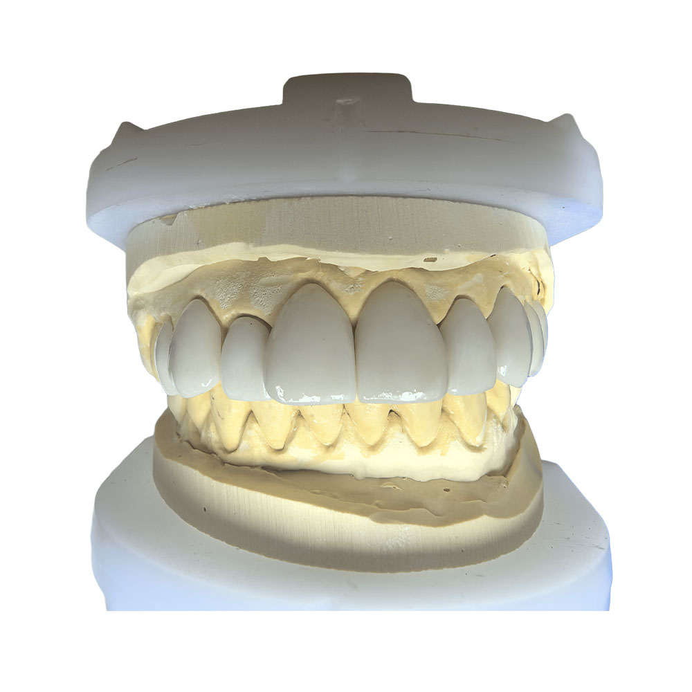
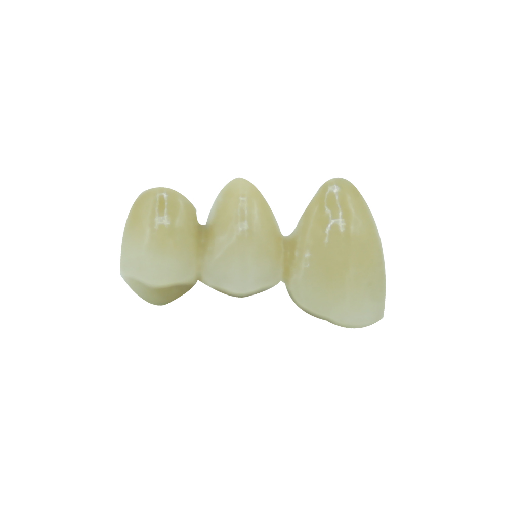
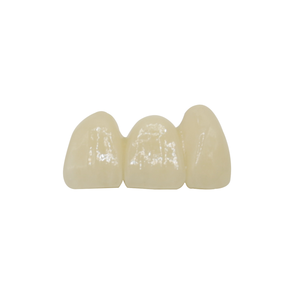
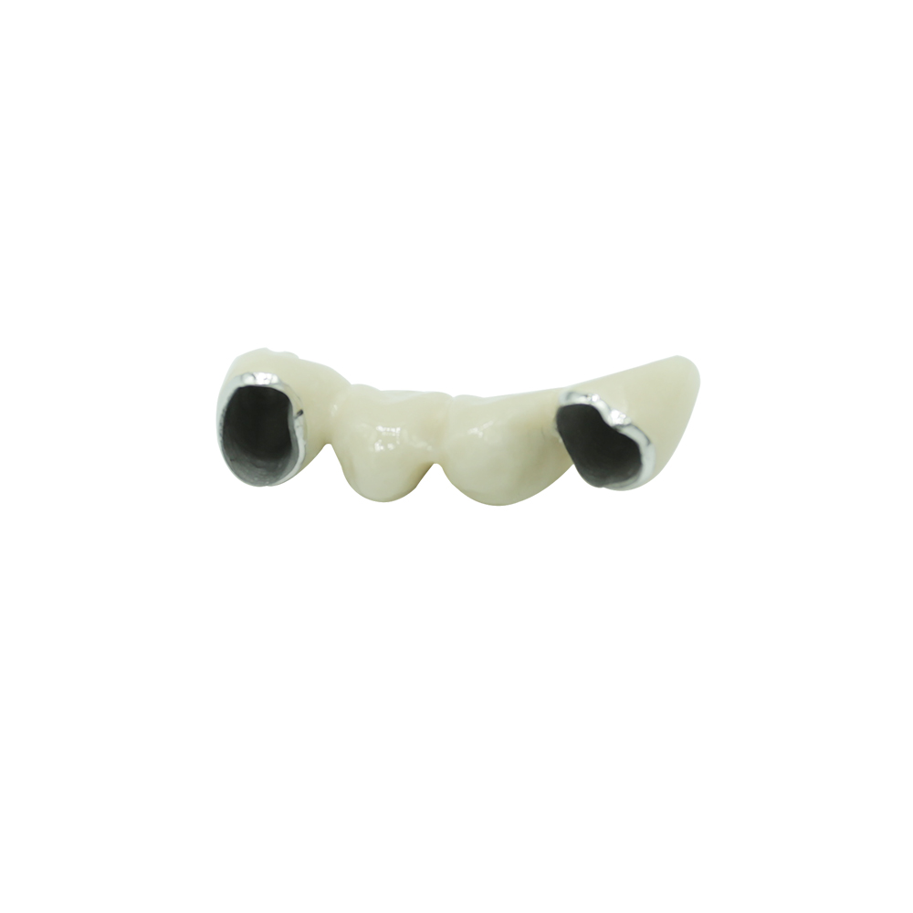
The patient presented with a failing 4-unit posterior dental bridge. The existing PFM bridge had chipped twice within 18 months — first along the occlusal surface of the second molar pontic, then at the distal cusp of the premolar retainer. Both repairs held less than six months.
The patient had a documented nickel sensitivity and visible gingival recession on the mesial abutment, where a thin gray line was becoming apparent at the margin.
The doctor’s referral noted: “Patient is frustrated. Wants to know if zirconia is the only option or if we can make PFM work this time.”
Why This Case Matters
Most discussions about pfm bridge failures stop at “porcelain chips sometimes.” But when a restoration fails twice in the same mouth, the question isn’t 무엇 failed — it’s why the design or execution allowed it to fail.
This case gave us a chance to demonstrate what a lab-side review should look like: identifying the mechanical cause, proposing a fabrication adjustment, and documenting the rationale so the clinician can explain it to the patient.
Four Obstacles the Doctor Flagged
O1: Insufficient Occlusal Clearance Led to Thin Porcelain
The original case photos showed <1.2 mm clearance on the molar pontic. When we model a pfm fixed bridge with that constraint, the metal framework consumes 0.4–0.5 mm, leaving ~0.7 mm for the porcelain veneer.
Research shows that porcelain fused to metal requires adequate thickness to withstand masticatory forces. When veneer thickness falls below 1.0 mm on load-bearing surfaces, the porcelain lacks structural support and becomes vulnerable to fracture under normal occlusal stress.
The doctor’s concern: “If I prep more, I’m into dentin sensitivity territory. If I don’t, we’re back to square one.”
O2: Patient Believes “PFM = Cheap and Outdated”
The patient had read that zirconia bridges are stronger and more durable than older PFM restorations. The question was framed as: “Why would I pay for another metal bridge when everyone says zirconia is better?”
The doctor’s concern: This wasn’t a clinical question — it was a trust question. The patient needed to understand 왜 PFM was still the right choice for this specific case, not just hear “it’s fine.”
O3: Metal Show-Through at Gingival Margin
The mesial abutment had 2 mm of recession since the original bridge was placed. The metal collar was now visible, creating a dark line the patient described as “looking like a cavity.”
The doctor’s concern: “I can’t promise the tissue won’t recede more. But I also can’t put an all-ceramic margin on a PFM bridge and expect it to survive five years in the posterior.”
O4: Nickel Sensitivity Required Alloy Verification
The patient’s chart documented contact dermatitis attributed to nickel jewelry. Studies indicate that base metal alloys used in some PFM restorations may contain nickel, which can trigger localized reactions in sensitized individuals.
The doctor’s concern: “I need written confirmation of the alloy composition. I can’t rely on ‘probably nickel-free’ when the patient has a known sensitivity.”
Our Response: Five Technical Requirements
R1: Modified Framework Design with Increased Metal Substructure (Answers O1)
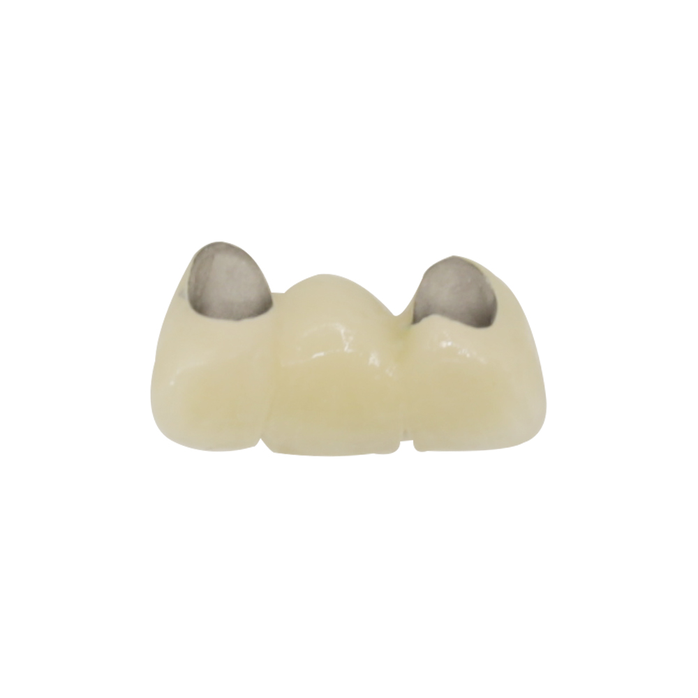
We proposed a thicker metal framework (0.6 mm instead of 0.4 mm) in the functional cusp areas, then reduced porcelain thickness to 0.8–0.9 mm on the occlusal surface.
This may sound counterintuitive — 덜 porcelain, not more — but the principle is material science: porcelain fused to metal derives its strength from the metal framework, not the veneer. A thin, well-supported veneer bonded to a rigid substructure will outperform a thicker veneer bonded to an under-dimensioned frame.
We documented the framework thickness on the digital design file and shared it with the doctor before milling, so the clearance question was settled 전에 casting.
R2: High-Noble Alloy with Full Material Traceability (Answers O4)
We fabricated the bridge using a palladium-silver high-noble alloy (noble metal content >60%) with zero nickel. The specific composition:
- Palladium: 52%
- Silver: 35%
- Gold: 8%
- Copper, tin, indium: balance
- Nickel: 0%
We provided a signed Material Safety Data Sheet (MSDS) and batch traceability certificate with the case delivery, so the doctor had documentation for the patient’s chart and could answer any future insurance or allergy-related questions.
Why high-noble instead of base metal? Two reasons. First, nickel-free base metal alloys (typically chromium-cobalt) still carry a small risk of sensitization in patients with known metal allergies. Second, high-noble alloys have a coefficient of thermal expansion closer to dental porcelain, which reduces the risk of porcelain delamination during the firing cycle — a factor that may have contributed to the original bridge’s repeated chipping.
R3: Porcelain Shoulder Margin on the Facial, Metal Collar on Lingual (Answers O3)
The mesial abutment received a modified design:
- Facial margin: porcelain shoulder extending 0.8 mm below the finish line, masking the metal and eliminating the visible gray line even if further recession occurs.
- Lingual margin: metal collar, which provides superior marginal seal and durability on the non-visible surface.
This isn’t a compromise — it’s an anatomy-specific design. Prosthodontic literature describes this “porcelain-facial, metal-lingual” approach as standard practice for anterior-facing surfaces on PFM restorations when esthetics and longevity must both be addressed.
The distal abutment retained a full metal collar because it was positioned outside the smile line and carried higher occlusal load.
R4: Written Comparison: When PFM Outperforms Zirconia (Answers O2)
We included a one-page technical note with the case delivery, written for the doctor to share with the patient. Key points:
Zirconia’s advantages:
- No metal substructure → no potential for metal show-through.
- Monolithic strength, especially in 3Y-TZP formulations.
- Biocompatible and metal-allergy-proof.
PFM’s advantages for this case:
- Lower cost for equivalent function (typically 30–40% less than multilayer zirconia).
- Proven long-term survival in posterior bridges; peer-reviewed studies report 10-year survival rates for PFM bridges in the 80–90% range when properly designed.
- Easier repair: porcelain chips on a pfm bridge can sometimes be polished or bonded chairside; fractured zirconia requires full replacement.
- Material flexibility: we could use a nickel-free high-noble alloy, which zirconia doesn’t require but also doesn’t offer as a variable.
The note made clear: zirconia isn’t “better” — it’s different. For this patient’s anatomy, occlusion, and material sensitivity, a properly designed PFM bridge offered the best balance of cost, function, and longevity.
R5: Pre-Delivery Digital Approval with Margin and Framework Views (Answers O1, O3, and Trust)
We sent the doctor three views before final glazing:
- Sectional view of the framework showing metal thickness at functional cusps.
- Margin close-up (facial side) showing the porcelain shoulder extension.
- Occlusal view showing the reduced-porcelain design and framework support.
This let the doctor verify that the clearance issue was solved and the esthetic concern was addressed 전에 we completed the case. If something looked off, we could adjust the biscuit-stage restoration — not after final glaze, and definitely not after try-in.
The Result
The bridge was delivered, seated, and adjusted in one appointment. No post-delivery chipping reported at the 6-month, 12-month, or 18-month follow-up.
The patient’s feedback (via the doctor): “It doesn’t look like a metal bridge, and it hasn’t chipped. I don’t know what they did differently, but it’s working.”
What We Actually Did Differently
Three things:
- We addressed the occlusal clearance problem at the design stage — not by asking for more prep (which wasn’t an option), but by redistributing material: more metal, less porcelain, better support.
- We gave the doctor technical documentation (alloy composition, margin design rationale, framework thickness) so the patient’s questions could be answered with data, not reassurance.
- We treated the case as an engineering problem, not a material-selection problem. The first bridge didn’t fail because PFM is inferior to zirconia. It failed because the framework was under-designed for the available clearance, and possibly because the alloy-porcelain thermal expansion mismatch contributed to delamination. Switching to zirconia would have solved the symptom (no porcelain to chip), but it wouldn’t have solved the 원인 (insufficient structural design for the given anatomy).
Why Labs Should Write Cases Like This
Most case studies in dentistry are written by clinicians or product manufacturers. That’s fine, but it leaves a gap: who documents what happens in the lab, and why the fabrication decisions matter?
When a doctor sends us a case with a documented failure history, they’re not just asking us to “make another bridge.” They’re asking:
- What went wrong the first time?
- How will you prevent it from happening again?
- Can you explain it in a way I can share with the patient?
Technical Sidebar: The Three Knowledge Gaps We Fill
Most online content about PFM bridges is written for patients: “What is it, how much does it cost, will it hurt?” That’s useful, but it doesn’t help clinicians or labs solve fabrication problems.
We focus on three areas that almost no one else documents publicly:
1. Clearance-Thickness-Framework Relationship
그리고 minimum clearance for a functional pfm dental bridge isn’t a single number — it’s a design triangle:
- Clearance = space available after tooth prep
- Framework thickness = metal substructure required to resist flexure
- Porcelain thickness = veneer required for esthetics and occlusal durability
You can’t dictate all three. If clearance is fixed (which it usually is), you balance framework vs. porcelain. Most failures we see come from labs defaulting to “standard” framework thickness (0.3–0.4 mm) even when the case demands more metal and less porcelain.
2. Thermal Expansion Coefficient Matching
Porcelain and metal expand at different rates when heated. If the mismatch is too large, thermal stress builds up during the firing cycle, creating microcracks that propagate under occlusal load.
High-noble alloys (gold-palladium, palladium-silver) typically have thermal expansion coefficients in the range of 13.5–14.5 × 10⁻⁶/°C. Base metal alloys (nickel-chromium, cobalt-chromium) range from 13.8–14.2, but the presence of nickel can shift the value and introduce sensitization risk.
We match the porcelain system to the alloy — not the other way around — because the alloy composition is usually constrained by cost, biocompatibility, or the doctor’s specification, while porcelain systems offer more flexibility.
Does this matter clinically? Prosthodontic research suggests that thermal mismatch contributes to porcelain delamination and chipping, especially in bridges subjected to high occlusal forces. Controlling it doesn’t 보증 success, but ignoring it increases failure risk.
3. Margin Design as an Anatomy-Specific Variable
The textbook says: “PFM crowns and bridges may have metal collars at the gingival margin or porcelain margins depending on esthetic requirements.”
Fine, but how do you decide?
Our rule:
- Visible margins (anywhere in the smile zone or where recession is likely): porcelain shoulder or butt joint, extended 0.5–1.0 mm apical to the finish line.
- Non-visible margins (lingual surfaces, distal molars): metal collar, which provides superior seal and durability.
- High-load abutments (cantilever bridges, long-span pontics): metal collar on all margins, even if esthetics are slightly compromised, because marginal integrity matters more than color match when the restoration is holding up the occlusion.
The distinction matters because porcelain margins are more fragile than metal margins. If you put a porcelain margin on a high-stress abutment just because “it looks better,” you’re trading durability for esthetics — which is fine if the doctor and patient agree to that trade-off. But it should be a documented decision, not a default.
Material Specifications for This Case
| Component | 재료 | 참고 |
| Framework alloy | Palladium-silver high-noble (52% Pd, 35% Ag, 8% Au, balance Cu/Sn/In) | Zero nickel; batch-traceable MSDS provided |
| Porcelain system | Feldspathic layering porcelain, CTE 13.8–14.2 | Matched to alloy expansion coefficient |
| Margin design | Facial: 0.8 mm porcelain shoulder; Lingual: metal collar | Anatomy-specific; documented in design file |
| Framework thickness | 0.6 mm at functional cusps, 0.4 mm at non-load-bearing areas | Increased from standard 0.4 mm to support thinner porcelain |
| Porcelain thickness | 0.8–0.9 mm occlusal, 1.2 mm facial | Reduced on occlusal surface to accommodate increased framework |
| 완료 | Glazed porcelain (occlusal/facial); polished metal (lingual collar) | No stain added per doctor request |
Quality control steps documented:
- Pre-casting framework thickness verification (digital calipers, 3 points)
- Post-casting framework fit check on stone model (before porcelain application)
- Porcelain application in two layers (opaque + dentin/enamel)
- Margin continuity check under 3.5× loupe
- Occlusal contact verification with articulating film on articulator
References and Further Reading
The technical principles discussed in this case study are supported by published prosthodontic literature and clinical guidelines. For readers interested in the evidence base:
- Cleveland Clinic: Dental Bridges: Types & Who Needs Them — comprehensive patient education resource covering bridge materials, procedure, and longevity expectations.
- My Specialty Dentist: Dental Bridge Materials Compared — prosthodontist-authored comparison of PFM, zirconia, and other bridge materials with survival rate data.
- Real Dental Costs: Tooth Replacement Options 2026: Cost Comparison Guide — cost and insurance coverage overview for various restorative options including PFM bridges.
- Veenstra Dental: Crowns & Bridges: The Art of Rebuilding Broken Smiles — clinical perspective on restoration design and material selection.
About ASD Dental Lab: We’re a full-service manufacturing facility (not a trading company) with in-house CAD design, metal casting, porcelain work, and quality control. Founded in 2012, we serve dental labs, clinics, and dentists in the US and Europe. Our Ceramic & Metal Department handles all PFM fabrication internally, which gives us direct control over framework design, alloy selection, and margin specifications.