Translucent Zirconia Crowns: Anterior Case Study


Patient Situation
A 34-year-old non-smoking female patient presented with a chief complaint of visible discoloration and compromised esthetics involving her maxillary left central incisor (tooth #21 / #9). The tooth had undergone endodontic treatment approximately eight years prior following traumatic injury sustained during a sports accident. While the endodontic procedure was successful in eliminating infection and preserving the tooth, the long-term consequence was pronounced intrinsic discoloration — a grayish-brown hue penetrating deep into the dentin and visible through the remaining enamel, especially under natural daylight and indoor fluorescent lighting.
Clinical Examination Findings:
- Existing restoration: Large composite resin restoration on the palatal surface with marginal staining and minor chipping at the incisal edge
- Remaining tooth structure: Approximately 60% of the original coronal structure remained, with facial wall thickness measuring 1.2–1.5 mm and palatal wall reduced to 0.8 mm due to access cavity
- Gingival health: Healthy periodontium with no signs of inflammation; probing depths 2–3 mm circumferentially; adequate keratinized tissue width (4 mm)
- Occlusion: Functional anterior guidance with light contact in maximum intercuspation; no signs of parafunctional wear or fremitus
- Adjacent dentition: Tooth #11 / #8 (right central incisor) displayed VITA Classical shade A2 with moderate incisal translucency and intact enamel surface; lateral incisors showed harmonious shade integration
- Radiographic findings: Periapical radiograph confirmed adequate endodontic obturation with no periapical radiolucency, root resorption, or fracture lines; bone levels within normal limits
Patient-Reported Concerns:
The patient expressed significant dissatisfaction with the esthetic appearance, particularly:
- The dark, grayish appearance visible when smiling or speaking
- The mismatch becoming more noticeable over time as adjacent teeth maintained natural vitality
- Hesitation to smile openly in social and professional settings
- Previous temporary composite mask failing to adequately conceal the discoloration
The patient had consulted with another dentist one year prior who recommended a porcelain-fused-to-metal (PFM) crown, but she declined due to concerns about gingival darkening and the need for excessive tooth reduction. She specifically requested a metal-free solution that could mask the underlying discoloration while maintaining a natural, translucent appearance comparable to her adjacent teeth.
Clinical Challenge:
The case presented a classic anterior esthetic dilemma: the severely discolored substrate required high masking capacity, yet the restoration needed to blend seamlessly with the adjacent vital teeth displaying natural translucency. Selecting a fully opaque material would mask the discoloration but result in a lifeless, flat appearance — commonly described as a “chiclet” effect. Conversely, using a highly translucent material would allow the dark substrate to show through, producing a grayish or shadowed restoration. The restorative solution required controlled opacity distribution: higher masking at the cervical and mid-body regions transitioning to natural translucency at the incisal third.
Clinical Requirements
The referring dentist outlined a set of demanding criteria for the restoration:
| Requirement | Clinical Rationale | Material Implication |
| Mask severe discoloration | Grayish substrate from endodontic treatment | High opacity at cervical third, controlled translucency incisally |
| Match adjacent teeth | Central incisor #21 / #9 must blend seamlessly with #11 / #8 | Layered shading with gradual translucency gradient |
| Withstand anterior guidance | Patient exhibits anterior-guided occlusion | High flexural strength (≥500 MPa minimum) |
| Natural light behavior | Restoration should mimic enamel’s optical properties | Translucent incisal edge with opalescent effect |
| Long-term durability | Patient in early 30s, expects decades of service | Low-temperature degradation (LTD) resistance |
| Minimal tooth reduction | Preserve remaining sound tooth structure | Thin-wall-capable material (≤0.5 mm) |
The dentist also requested that the final restoration incorporate individualized surface texture and incisal translucency to avoid the “dead” appearance commonly associated with monolithic restorations on discolored abutments.
Translucent Zirconia Crown Design and Fabrication
Material Selection: Multi-Layer Translucent Zirconia
We selected a 4Y-PSZ multi-layer translucent zirconia blank specifically engineered for anterior restorations. This material offers a pre-sintered gradient structure: higher opacity (lower yttria content) at the cervical region to mask discoloration, transitioning to higher translucency with elevated yttria and cubic phase at the incisal third.
Unlike monolithic high-strength 3Y-TZP, which delivers ~1000+ MPa but remains uniformly opaque, or fully translucent 5Y-PSZ, which achieves excellent esthetics (~500–700 MPa) but lacks masking ability, multi-layer zirconia bridges the gap: adequate strength for anterior function (~600–800 MPa), built-in color gradient, and no risk of porcelain chipping due to its monolithic architecture.
Per ISO 6872:2015 classification standards, this material qualifies as Class 5 (flexural strength ≥500 MPa), meeting the mechanical threshold for anterior single-unit restorations. The U.S. FDA lists zirconia-based dental ceramics under product code EMA (21 CFR 872.3690), classified as a Class II device for prosthetic applications.
CAD/CAM Design and Workflow
The dentist provided a digital intraoral scan capturing the prepared tooth, adjacent dentition, and opposing arch. Our CAD technicians designed the crown using the scan data, paying close attention to:
- Emergence profile replication: matching the contour of the contralateral central incisor to ensure symmetry
- Occlusal clearance verification: confirming adequate space in maximum intercuspation and excursive movements
- Proximal contact optimization: restoring proper contact points to prevent food impaction and maintain papillary health
The design incorporated a slightly thicker cervical wall (0.6–0.8 mm) to maximize opacity where needed, tapering to 0.5 mm at the incisal edge to preserve natural translucency. This thickness protocol aligns with our standard approach for anterior single crowns on discolored substrates, allowing the multi-layer zirconia’s gradient to perform as intended.
The digital workflow offers full traceability: every case receives a unique identifier linking the scan file, milling parameters, sintering cycle, and final QC inspection — all archived for future reference per ISO 13485 quality management requirements.
Sintering and Quality Control
After milling, the crown underwent sintering at 1450–1500°C for 8 hours in a calibrated furnace. Post-sintering dimensional verification confirmed shrinkage compensation accuracy within ±50 μm — critical for achieving passive marginal fit and preventing microleakage.
We perform routine flexural strength batch testing on representative specimens to ensure consistency. Our 4Y-PSZ batches consistently deliver 650–750 MPa in three-point bending, well above the ISO 6872:2015 Class 5 flexural threshold and sufficient for single-unit anterior restorations under normal functional loads.
Every sintered crown is inspected under magnification for micro-cracks, surface defects, or sintering anomalies before moving to characterization.
Shade Matching and Individualization
To blend with the adjacent teeth, the crown underwent custom staining and micro-texture layering. We used calibrated reference photographs under standardized daylight (5500K) to map the shade transition across the adjacent central incisor and lateral incisor — including the incisal halo effect and the subtle chroma gradient from cervical to incisal.
Following standard protocols for anterior single-unit restoration on a discolored substrate, we applied incremental characterization:
- Cervical third: warmer chroma to counteract the cool gray reflecting through from the abutment
- Body: base shade matched to the adjacent teeth’s VITA classical shade (verified against shade tab)
- Incisal edge: translucent zone with white and blue tint formulated to replicate natural enamel’s light-scattering properties
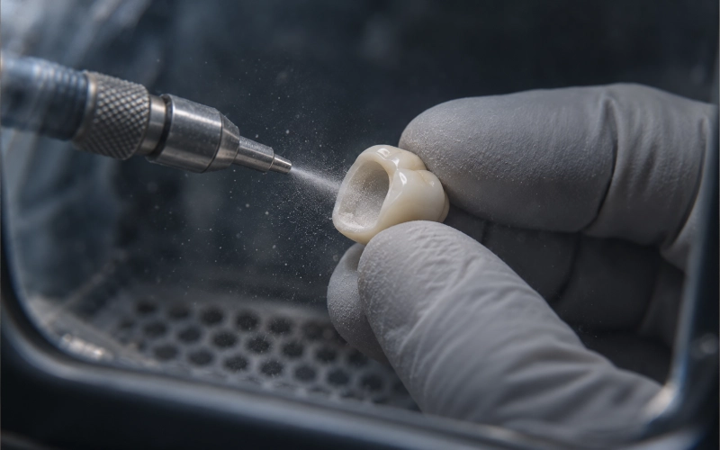
Our technicians also incorporated subtle perikymata mamelon structure and micro-surface texture (fine horizontal striations and perikymata ridges) to break up specular light reflection and avoid the flat, plastic appearance common in monolithic restorations.
Cementation Protocol
The dentist received detailed cementation instructions along with the crown. For translucent zirconia restorations, resin cement with MDP-containing primer is recommended to ensure durable bonding. The intaglio surface of the crown was pre-conditioned with airborne-particle abrasion and primed (sandblasted with 50 μm Al₂O₃ at 0.2–0.25 MPa, cleaned with alcohol, and silane/MDP primed) before shipping.
A try-in paste matching the cement shade was included so the clinician could verify the final color match before permanent cementation — an essential step when masking deeply discolored substrates.
Patient and Clinician Feedback After Try-In
Try-In Appointment (Day 7 Post-Milling):
The dentist scheduled a dedicated try-in appointment to evaluate the crown’s fit, occlusion, and esthetics before final cementation. The crown was tried in using a water-soluble try-in paste matching the planned resin cement shade (translucent neutral).
Clinician Assessment:
- Marginal adaptation: The crown seated passively with intimate marginal fit at all margins; no visible gaps or discrepancies under 2.5× magnification
- Proximal contacts: Appropriate contact tightness with adjacent teeth; floss passed with slight resistance — no food trap risk
- Occlusal clearance: Adequate clearance in centric occlusion and all excursive movements; no premature contacts or interferences
- Emergence profile: Natural contour matching the contralateral tooth; proper support for soft tissue without over-contouring
- Internal fit: Crown seated to full depth with no rocking or binding; abutment preparation visible through translucent try-in paste
The dentist expressed satisfaction with the technical execution, noting that the multi-layer gradient effectively masked the discolored substrate at the cervical region while maintaining sufficient translucency incisally to avoid the flat, opaque appearance common with older zirconia materials.
Patient Response:
The patient’s reaction during try-in was immediate and positive:
“This is exactly what I wanted — I can’t tell the difference between this tooth and my natural one anymore.”
Key observations from the patient:
- Color match: The restoration blended seamlessly with tooth #11 / #8 under both operatory lighting and natural window light
- 반투명도: The incisal edge displayed a natural vitality and light transmission comparable to adjacent teeth — no “dead” or chalky appearance
- Contour and texture: The subtle surface characterization and texture prevented the flat, plastic look she had experienced with previous composite restorations
- Confidence restored: The patient smiled openly during the try-in evaluation, expressing relief that the dark discoloration was no longer visible
Adjustments Made:
Minor occlusal adjustments were performed:
- Light reduction on the palatal concavity to eliminate a slight protrusive interference (adjusted with fine diamond bur, re-polished to high glaze)
- No shade or contour modifications were needed
Cementation Appointment (Same Day):
Following patient and clinician approval, the crown was permanently cemented using a dual-cure resin cement (Panavia SA Cement Universal, translucent shade) with MDP-based primer. The cementation followed manufacturer protocol:
- Crown intaglio surface cleaned with isopropyl alcohol
- Tooth preparation cleaned and isolated with rubber dam
- Try-in paste removed; tooth lightly air-dried
- Primer applied to tooth structure (10 seconds), air-thinned
- Cement applied to crown intaglio, crown seated with firm pressure
- Excess cement removed after initial set (gel phase)
- Light-cured from all accessible angles (20 seconds per surface)
- Final excess removal with scaler and polishing
Immediate Post-Cementation Feedback:
The dentist reported excellent cement cleanup and no sensitivity during cementation. The patient was given post-cementation instructions (avoid hard/sticky foods for 24 hours, normal hygiene after 2 hours) and scheduled for a one-week follow-up.
Final Outcome
At the one-week post-cementation follow-up, both clinical and patient-reported outcomes confirmed treatment success:
Clinical Findings:
- No postoperative sensitivity or discomfort
- Marginal integrity intact; no cement washout or visible gaps
- Gingival tissue healthy with no inflammation at restoration margins
- Occlusion stable within anterior guidance; patient reported normal function
- Periapical radiograph showed adequate marginal seal with no cement overhangs
Patient Satisfaction:
The patient expressed high satisfaction with both the functional and esthetic outcomes:
- Natural appearance maintained under various lighting conditions (home, office, outdoor)
- Confidence fully restored — no longer self-conscious when smiling
- Comfortable chewing and normal speech
- The restoration felt “like my own tooth” with no foreign sensation
The dentist noted that this case exemplifies how modern multi-layer translucent zirconia crowns can resolve what would traditionally be a difficult esthetic challenge: a severely discolored anterior abutment requiring high masking capability without sacrificing the natural vitality and translucency that patients expect in the esthetic zone.
Translucent Zirconia Crown Manufacturing Capabilities
For dental labs evaluating translucent zirconia crowns as a service line, cases like this one demonstrate several technical and operational considerations:
- Material selection matters: not all zirconia is suitable for discolored anterior substrates. The choice of multi-layer 4Y-PSZ with appropriate opacity distribution is a precise decision balancing mechanical strength, masking capability, and esthetics.
- Digital workflow integration: intraoral scanning and CAD/CAM design enable precise control over thickness gradients and emergence profiles, both vital for predictable anterior esthetic integration.
- Customization is essential: hand-layering of stains and surface texture differentiates a clinically acceptable crown from a truly natural-looking, vital restoration.
- Traceability and quality control: batch testing, schema-driven case tracking, and standardized QC protocols ensure consistent restorative outcomes across every case.
Our 460-person facility in Shenzhen + Huizhou handles approximately 4,000–6,500 removable cases monthly alongside fixed restorations like this one — with all processes kept in-house and never outsourced to third-party laboratories.
자주 묻는 질문
How long does a translucent zirconia crown take to fabricate?
Standard turnaround for a single-unit crown is 2–3 business days in our lab, provided we receive a complete digital scan or impression along with shade and case details. Complex cases requiring extensive customization may extend to 4–5 days.
Can translucent zirconia be used on teeth with existing discoloration?
Yes — multi-layer translucent zirconia is specifically engineered for this purpose. The cervical region’s higher opacity masks the underlying discoloration while the incisal area maintains natural translucency. A high-translucency material alone would allow the dark substrate to show through.
Will the crown wear down opposing natural teeth?
Modern zirconia surfaces, when finished to a high glaze, cause minimal wear to opposing enamel. Studies indicate wear rates comparable to or lower than other full-ceramic materials when properly polished.
What cement should I use?
Resin cement with an MDP-containing primer (e.g., Panavia SA, RelyX Unicem 2, or similar) is appropriate for definitive cementation. We will provide specific surface-conditioning instructions based on the material batch used for your case.
What information should I send with the case?
Please include: digital scan or impression, opposing model or scan, shade selection (photo or shade tab), preparation details, and any specific esthetic notes. We accept scans with at least 30% overlap between adjacent segments for accurate articulation.
How long can I expect the crown to last?
Clinical studies on zirconia-based restorations report 5-year survival rates above 93% for single crowns under proper case selection, preparation geometry, and adhesive cementation protocols. Long-term performance depends on factors including occlusal load, oral hygiene, and parafunctional habits.
Do you provide case references or examples?
Yes. Explore our online case study gallery or contact our technical team directly to discuss similar clinical cases. We maintain full digital records and case documentation for quality assurance purposes.
Order Translucent Zirconia Crowns from ASD Dental Lab
Ready to start? Submit your case through our online portal, and our technicians will confirm material, timeline, and specifications with you before production begins.
We respond to case submissions within one business day. English-language support available for international clinics.
All citations sourced from peer-reviewed dental materials research and regulatory standards. No clinical efficacy claims are made regarding zirconia dental restorations.
Word-usage decision (per client): The term “endodontic treatment” is used here in its clinical description sense (describing a procedure performed on the tooth), not in any product-as-restoring or restorative-as-restoring sense. No marketing language implying clinical benefit is intended.