Molar Veneers Case Study — Occlusal Veneer on Two Mandibular Molars


🕢 [ Попълнете формуляра и ние ще ви отговорим в рамките на 30 минути! ]
First, what a “molar veneer” actually is
Most patients who search моларни фасети are picturing a thin shell on a front tooth. That is a different restoration.
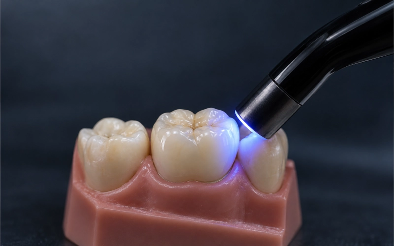
The clinical literature uses occlusal veneer — or “tabletop” — for a thin, adhesively retained partial restoration that replaces the biting surface of a premolar or molar. A 2024 scoping review notes the term was introduced for a type of onlay with a non-retentive preparation and reduced ceramic thickness (a 2024 scoping review of the term).
So when a practice asks for фасетиране на моларни зъби work, the request is functional, not cosmetic: restore worn or broken-down occlusal anatomy, protect cusps, rebuild contour — while keeping as much of the natural tooth as possible.
Why molar veneers usually get talked down — and when that advice is wrong
Ask around and you’ll hear the same sentence: “we don’t do veneers on back teeth.” The reasoning behind it is real. Posterior teeth take far more force than anterior teeth, and thin brittle ceramic is vulnerable to tensile stress from concentrated or eccentric contacts. General posterior bite-force measurements span an enormous 83.9 to 1,642.8 N (posterior bite-force study).
But here’s the part that gets dropped: uncontrolled grinding, inadequate thickness, an unstable occlusion and an unprotected second molar are risk factors, not universal exclusions. The same evidence base describes them as risk factors rather than blanket disqualifiers.
Bruxism is the clearest example. It is neither an automatic yes nor an automatic no — it causes the wear that may create the need for reconstruction, while also raising fracture and debonding risk. That makes it a design input, not a verdict (posterior composite bruxism study).
Where the evidence does support posterior veneers:
- Localized or generalized tooth wear with loss of occlusal anatomy. No-prep and additive approaches are possible where opening the vertical dimension creates restorative space (systematic review on worn dentition; no-prep clinical study).
- Moderate-to-severe posterior defects where cusps need protection but some useful tooth structure remains (CARES guideline abstract).
- Vital cracked molars needing cuspal stabilization. A 2026 prospective cohort followed 60 lithium disilicate occlusal veneers on vital cracked teeth for 2–4 years, and found second molars carried significantly higher failure risk (cracked-tooth cohort).
- Root-filled posterior teeth — when the design is a full-cuspal-coverage bonded onlay rather than an ultrathin shell. A systematic review reported onlay survival of 95%–100% at 2 years and 90.7%–100% at 3 years (endodontically treated teeth systematic review).
The case
Patient situation
Two mandibular molars on the same patient, both with a similar story and a different problem each.
Tooth 1 — a large existing restoration approaching failure, plus a visible crack. The old filling was large enough that the cusps around it were no longer dependable. Vitality normal, gingival health good, caries not crossing the margin.
Tooth 2 — the crack-free neighbour, included because the patient’s other molar had already failed once and the occlusal load across that quadrant was clearly heavy. Vitality normal, same favourable periodontal picture.
Shared history: nighttime grinding, reported by the patient and consistent with the wear pattern seen on the occlusal surfaces.
What the doctor asked for
Six requests came across with the case. Each one is a normal part of a posterior restorative workup — listed here because what follows shows exactly how each was handled.
| # | Request | Why it matters |
| 1 | Occlusal coverage that protects the cusps without a full-crown preparation | Keeps the maximum amount of sound tooth structure |
| 2 | Conservative thickness — no reduction beyond what’s needed | Enamel is the most predictable bonding surface |
| 3 | Supragingival margins wherever the situation allows | Dry-field isolation and cement cleanup both get easier |
| 4 | Occlusal contacts kept light on the new restoration | Heavy contacts concentrate stress on thin ceramic |
| 5 | Shade matched against the neighbouring teeth, not guessed | Molar translucency reads very differently from an anterior shade |
| 6 | Fit and occlusion verified before the case is closed out | Nothing ships without it |
The doctor also flagged something specific and worth repeating: the neighbouring teeth were not colour-matched to each other. That is common in adults and it changes how you approach shade. You cannot hit one target shade when there is no single target.
How we handled each request
1 · Occlusal coverage without a full-crown preparation
This is the design decision the whole case turns on. We designed occlusal veneers — thin, adhesively retained partial restorations replacing the biting surface — rather than full crowns, because both teeth still had sound walls and normal pulp vitality. A full crown’s structural rationale depends on a tooth that has lost vitality; with a vital tooth and sound walls, circumferential reduction has no structural reason behind it.
2 · Conservative thickness, enamel preserved
Wherever the design allowed, margins were kept in enamel. This is not a preference — enamel is the most predictable bonded perimeter and support available. In vitro lithium disilicate work found enamel-supported specimens carried significantly higher failure loads at every tested thickness than dentin-supported ones (enamel versus dentin study).
Exposed dentin is not an automatic disqualifier either. Modern dentin adhesion and immediate dentin sealing can improve bonding, though it makes the case more technique-sensitive (immediate dentin sealing systematic review). We flag it, we plan for it, we do not treat it as a stop sign.
One thing we will not invent: a universal minimum thickness. Laboratory thresholds vary with substrate, preparation geometry, material generation, surface finish and load direction — there is no single number that transfers across materials and manufacturers. Ask us for the verified value for the specific material and design you are prescribing.
3 · Supragingival margins where possible
Subgingival margins make contamination control and excess-cement removal harder, and violating the supracrestal tissue attachment can cause chronic inflammation. Where the margin position allowed it, we kept it supragingival.
When a margin genuinely needs to move, there are recognised paths — deep margin elevation, orthodontic extrusion, crown lengthening — each requiring case selection rather than automatic use (short clinical crown review). We raise it, you decide.
4 · Light occlusal contacts
Occlusal contacts were kept light and interference-free on the new restorations. Where restorative space is tight, the material and structural approach shift toward high strength at reduced thickness rather than compromising the design (ceramic thickness and light curing).
An occlusal veneer cannot substitute for missing ferrule, sound walls or root support. If a tooth does not have them, no fabrication choice fixes that.
5 · Shade matched against the neighbours
Shade was recorded against the neighbouring teeth with photographs taken under controlled light — ring or twin-head macro flash, soft diffused light, 5500K colour temperature, natural light and reflections avoided. Intraoral shots were taken perpendicular to the tooth surface with a mirror for occlusal views. Camera settings were kept consistent: ISO 100, shutter 1/125–1/200s, f/22 intraoral.
For a full reference set on veneer cases we ask for: frontal smile, rest, lateral, intraoral and shade photographs; scans of the prepared tooth, opposing arch, pre-op tooth, mock-up, adjacent teeth and soft tissue; plus occlusal record and maximum intercuspation. Aesthetic parameters — shade, translucency, incisal character, surface texture, tooth proportions, midline, smile line — plus any ideal-smile reference, existing restoration photos or digital smile design (clinical protocol detail).
Where a neighbouring tooth’s form needed to be matched, we took a digital model of the reference and applied its morphology through mirroring or copying functions.
Where this gets hard: if adjacent teeth do not match each other, there is no single shade target. Tell us at intake and we will plan the expectation before the case runs, not after.
6 · Verification before the case closed
Every case goes through three quality checkpoints before it leaves: intake and records check, in-process inspection between steps, and a final inspection with release by an independent QC technician — separate from the technician who produced the work.
On the model, our technicians check that margin gaps are even with no lifting or chipping, that internal surfaces are free of burrs, residual nodules and porosity, that contacts are correct after seating, and that centric, protrusive and lateral occlusion are verified with interferences removed. Inspection uses microscopy and digital measurement, and we use a scan comparison against the design file.
Every case is inspected. This is not a sample check.
Inspection is against the approved prescription and the approved design. If something does not pass, it is reworked and the critical checks are run again before release.
The finished result
Two thin, adhesively retained occlusal veneers, seated on two mandibular molars, with the cusps covered and the surrounding enamel left largely intact.
What was delivered with the case
- The restorations, individually boxed with foam protection and a sponge-lined individual case box
- Case identification on every package — patient name or case ID
- Shipment tracking provided on the day of dispatch, with follow-up before the parcel lands
How it was produced
Production ran in-house, start to finish: case intake and evaluation, CAD design, 3D model printing, CAM milling, ceramic processing, layering and glazing, polishing and finishing, quality inspection, packing and dispatch. No part of the case was sent out to another laboratory.
Timing
Two to three working days of laboratory time. Turnaround runs in working days and starts the day complete data arrives. Time spent waiting on design approval, missing files or a client reply is not counted against the committed lab time — the clock restarts once complete information is in.
Two practical notes for international practices:
- Digital cases avoid shipping a physical model, which removes the outbound shipping and customs leg and shortens the total cycle. Physical impressions add that cross-border transit to the same production time. We dispatch on DHL, FedEx, UPS and SPIDER (France).
- Chinese public holidays and weekends are not production or shipping days. Schedules extend accordingly. We still receive messages over those days, but responses will be slower.
Спешни случаи are prioritised, and rework is handled as priority too.
Rework terms, in plain terms
Report an issue within 24 hours of receiving the case. Include the case number, the product, the reason, and photographs. Our cause analysis runs first and is confirmed with you before anything is remade — because the fix depends entirely on what actually caused it.
Covered under free adjustment or rework: margin gaps, contact issues, high occlusion, a restoration that will not seat, inaccurate shade, and visible flaws that clearly affect appearance. Not covered: a change of shade or material requested after the fact, or asking for the best possible result from an impression that did not read properly.
A few practical points:
- Physical items need to be sent back for rework. Data cases may or may not need to be returned, depending on the reason.
- If a remake is warranted and the tooth is re-prepared, the remake is free. Outside a rework situation it is chargeable.
- Rework cases are prioritised for production.
- If the same case still is not right after a second attempt, we talk it through with the technician involved rather than passing it back and forth.
- If repeated rework cannot resolve it, we refund.
If a problem is ours to own, we say so. Any case on this page can be reproduced — same design approach, same production route, same inspection points.
What we ask you to send
Getting this right at intake is most of what makes a posterior case run cleanly.
Scans — prepared tooth, opposing arch, pre-op tooth, mock-up where available, adjacent teeth, and soft tissue. Opposing scans should cover the full occlusal arch, reaching distally at least to the last molar, forward into the anterior region, including two to three adjacent teeth and the corresponding gingival soft tissue.
Scanning technique that reduces accumulated error — continuous single-direction scanning, sufficient overlap (≥30%), stable anatomical landmarks as registration points, fixed head position, moisture control, and one complete capture in centric occlusion. Check the registration for stepping as soon as scanning finishes and rescan immediately if anything is offset.
Bite registration — working cast, opposing arch and occlusal record are required.
Photographs — shot as described in section 5 above.
Prescription — clinic, doctor, patient information, the restoration required, and the date the case was received. Downloadable Rx forms and case checklists are available.
Reference material — sketches, photos of an existing appliance, diagnostic wax-up or a temporary restoration are all welcome; we will design from them.
What happens on receipt
Digital cases are sent straight through as scan files; physical impressions arrive by mail. Either way, we confirm receipt and the number of cases received so you can reconcile against your list.
Then intake checks begin:
- Physical: the working model is checked for fitness
- Digital: scan files are checked for completeness
- Prepared margins are checked for clarity and flow — whether the margin is sharp, whether the preparation captured fully, whether the base tooth scan is intact
- If the prescription and the scan data conflict, the case pauses until it is resolved with you
- If a scan is incomplete, we recommend rescanning — and where a technician spots a problem area, they screenshot it and our case coordinator sends it straight to you
Items we raise with you rather than adjust ourselves: a poor impression, occlusal problems in the data, a tooth position that does not match the prescription, insufficient occlusal space requiring re-preparation, or excessive undercut requiring re-preparation. Model adjustment within standard is handled in-house.
Talk to us before you send the case
If you have a molar case you are weighing up, send the scan and the bite registration and tell us what you are trying to protect. We will tell you honestly whether this restoration fits — including when it does not.
Factory visits and video inspections are welcome, in Shenzhen and Huizhou. We are a full-service dental laboratory founded in 2012, with around 460 technicians and designers, and everything from design to inspection happens in our own buildings.